 2026, Vol. 46


放射性肺损伤(RILI)是胸部恶性肿瘤放射治疗时常见的并发症,其发生率高达5%~20%[1]。根据临床特征可分为两个阶段:早期表现为放射性肺炎(RP),出现在放疗后的1~3个月;中晚期发展为不可逆的放射性肺纤维化(RIPF)[2],一般出现在放疗后的6~24个月。放射性肺纤维化一旦发生,可导致呼吸功能障碍,甚至呼吸衰竭乃至死亡。目前临床上针对放射性肺损伤的药物防治手段疗效有限,因此亟待深入阐明RILI的发病机制,进而寻求治疗RILI的新策略。
围绕RILI的致病机制的主流学说有细胞损伤学说和细胞因子学说[3-4]。近年研究表明,多种免疫细胞通过识别损伤相关分子模式(DAMP)形成复杂的交互调控网络,在RILI早期炎症反应阶段到纤维化重塑阶段均发挥重要调控作用[5]。例如,T细胞亚群平衡决定炎症状态[6];M1型巨噬细胞和M2型巨噬细胞分别参与放射性肺炎和纤维化的发展[7];中性粒细胞释放活性氧等物质加剧炎症[8]。此外,免疫细胞间通过细胞因子介导互作,如Treg细胞通过释放抗炎细胞因子促进巨噬细胞向M2型极化[9],Th17分泌IL-17促进中性粒细胞募集,调控肺实质细胞功能,共同推动RILI的炎症与纤维化进程[10]。本文对免疫细胞及其与肺组织细胞间在RILI中的互作网络机制进行了总结和分析,旨在为提高临床RILI的防治水平提供必要的理论研究依据。
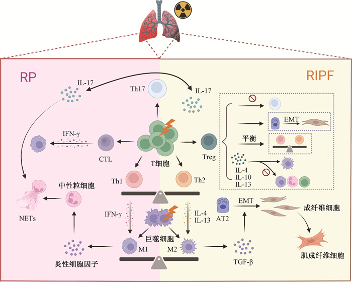
一、电离辐射诱导肺损伤过程中免疫细胞交互网络的细胞构成正常肺组织中的免疫细胞通过识别和清除病原体、调节炎症反应以及参与免疫记忆的形成保护肺部免受感染和损伤[11]。近年来研究表明,辐射通过改变肺部免疫微环境,形成复杂的免疫细胞调控网络,损害肺正常组织细胞,示于图 1[12-13]。因此,阐明RILI发生及进展过程中免疫微环境中各类细胞的功能变化机制对RILI的基础及临床研究均具有重要意义。
|
注:RP. 放射性肺炎;RIPF. 放射性肺纤维化;Th. 辅助性T细胞;CTL. 细胞毒性T细胞;Treg. 调节性T细胞;M1. M1型巨噬细胞;M2. M2型巨噬细胞;AT2. Ⅱ型肺泡上皮细胞;EMT. 上皮-间质转化;NETs. 中性粒细胞胞外陷阱;IL. 白细胞介素;IFN-γ. 干扰素γ;TGF-β. 转化生长因子β 图 1 放射性肺损伤中巨噬细胞、中性粒细胞以及T淋巴细胞间的互作网络 Figure 1 The interaction network among macrophages, neutrophils, and T lymphocytes in radiation-induced lung injury |
1.T细胞在RILI的免疫微环境中发挥核心调控作用
(1) Th细胞亚群之间的平衡决定RILI炎症状态:Th细胞亚群的极化和分布以及各亚群之间的平衡决定炎症状态[6],调控RILI的发生与进展。其中,Th1细胞分泌干扰素γ(IFN-γ)、白介素2(IL-2)等促炎因子,增强巨噬细胞和细胞毒性T细胞的活性,参与早期炎症反应,并发挥一定的抗纤维化作用[14-15]。Th2细胞分泌的IL-4、IL-13等细胞因子促进成纤维细胞活化和胶原蛋白沉积,在放射性肺纤维化进程中发挥关键作用[16]。Th17细胞产生IL-17家族细胞因子,主要作用于先天免疫细胞和上皮细胞等,促进中性粒细胞的生成和募集,调控上皮免疫屏障的完整性和防御功能[10]。同时,IL-17诱导成纤维细胞和肺上皮细胞中趋化因子表达,促进炎症进展[17]。此外,Paun等[15]研究发现经辐射处理的IL17-/-小鼠,因缺乏Th17细胞,而未发生肺炎和纤维化,提示Th17细胞与RILI纤维化密切相关。
(2) CTL通过多种机制参与RILI炎症的发展:CTL是适应性免疫的重要组成部分,其在清除细胞内病原体并介导长期免疫保护方面发挥关键作用。CTL细胞通过释放颗粒酶和穿孔素等细胞毒性分子,直接杀伤携带靶抗原的细胞,加剧肺组织的炎症反应。此外,CTL细胞分泌的IFN-γ和肿瘤坏死因子α(TNF-α)等细胞因子不仅可以增强局部炎症反应,还可通过激活巨噬细胞和中性粒细胞进一步放大组织损伤[18]。Yang等[19]研究报道,辐射暴露后,组织中CD8+ T细胞的浸润与CCL3水平的升高呈正相关,在缺乏CCL3或其受体CCR1的小鼠中发现RILI损伤减轻。因此,CTL在RILI过程中通过细胞毒作用和炎性因子释放加剧肺组织损伤。
(3) Treg在维持RILI免疫稳态中起关键作用:Treg细胞作为CD4+ T细胞的一个亚群,在RILI中通过多途径发挥免疫调节作用。在炎症发生时,Treg细胞通过分泌IL-10抑制中性粒细胞的渗出,减少IL-6、IFN-γ等促炎因子的分泌[20];通过促进中性粒细胞的凋亡,增强巨噬细胞对死亡中性粒细胞的吞噬作用;以及通过释放IL-4等抗炎细胞因子抑制单核细胞的活性,促进巨噬细胞向M2抗炎表型极化[9]这3个途径调节免疫反应。同时,Treg细胞通过分泌IL-10、转化生长因子β(TGF-β)和IL-35,抑制CD4+ 和CD8+ T细胞介导的炎症反应,促进炎症消退[21-22]。此外,Treg细胞还可以平衡Th1/Th17反应,刺激肺上皮细胞和内皮细胞的增殖[23]。在肺纤维化形成期,Treg细胞主要通过促进纤维细胞积累、促进β-连环蛋白(β-catenin)介导上皮-间质转化(EMT)、减弱Th17反应和调节Th1/Th2细胞因子平衡等,参与辐射诱导的肺纤维化[24-26]。由此可见,Treg细胞通过抑制炎症反应和参与纤维化过程在RILI中发挥双向调控作用。
2.巨噬细胞是参与RILI发生进展的关键调控环节:巨噬细胞以经典激活的M1型巨噬细胞(促炎)和替代活化的M2型巨噬细胞(抗炎/促纤维化)为代表,在RILI病变进程中起着重要作用[7]。
M1型巨噬细胞可被脂多糖(LPS)、IFN-γ等细胞因子激活,通过分泌包括TNF-α、IL-1β、IL-6等炎性细胞因子,促进放射性肺损伤早期阶段的炎症反应[27]。此外,电离辐射诱导肺巨噬细胞生成大量活性氧(ROS),由此触发ROS介导的氧化级联反应;而升高的ROS水平又能诱导巨噬细胞向M1型极化[28]。研究进一步表明,RILI中NLRP3炎性小体的活化、STING信号的激活及肺上皮细胞来源的CCL7等均可驱动巨噬细胞向M1极化,并通过NF-κB/STAT1等通路加重炎症[29-31]。随着病程进展,IL-4、IL-10等抗炎细胞因子的分泌及ROS的降低推动巨噬细胞向M2型极化[32-33]。M2型细胞通过释放TGF-β、血小板源性生长因子(PDGF)等促纤维化因子,诱导成纤维细胞分化为肌成纤维细胞,并激活Wnt/β-catenin通路,促进细胞外基质沉积,从而在放射性肺纤维化的发生与进展中起关键作用[34-35]。
3. 中性粒细胞通过直接参与和放大免疫反应加剧放射性肺炎:中性粒细胞是最早被募集到炎症部位的细胞之一。研究发现,肺组织发生炎症时中性粒细胞表面的G蛋白偶联受体可被局部驻留细胞产生的CXCL1和CXCL2等炎症介质结合并被激活,进而介导中性粒细胞的活化和募集[36]。
相关研究表明,中性粒细胞可通过吞噬作用、释放活性氧和弹性蛋白酶等直接损伤肺组织[8]。活化的中性粒细胞还能释放中性粒细胞胞外陷阱(NETs),导致免疫反应的放大,加剧肺上皮细胞和血管内皮细胞损伤[37]。此外,炎症部位的中性粒细胞通过释放多种信号介质,如细胞因子、细胞内颗粒、脂质及过氧化氢,调控各类免疫细胞的定向迁移[38]。同时,中性粒细胞还借助细胞间接触进行信息传递,影响并协调其他免疫细胞的功能活动,共同调节损伤区域的病理进程[39]。综上,中性粒细胞通过直接杀伤和免疫调控作用促进放射性肺炎的发展。
4. 其他免疫细胞在RILI发生发展中的功能调控: 除T细胞、巨噬细胞和中性粒细胞外,B细胞、NK细胞和树突状细胞(DCs)也可通过不同机制参与RILI的免疫调控。
B淋巴细胞在适应性免疫系统中起着至关重要的作用,研究显示其受体信号通路在RILI中受circRNA与miRNA的显著上调或下调而被异常调控[40]。此外,B细胞可以分化为调节性B细胞(Bregs)和效应性B细胞(Beffs)。Bregs可通过分泌抗炎细胞因子抑制炎症反应,在RILI中发挥保护作用[41-42]。目前对于RILI中B细胞的功能研究尚不全面,未来需进一步明确。NK细胞作为先天免疫细胞,既能够直接清除被感染的细胞,也可以通过分泌TNF-α、IFN-γ等细胞因子促进炎症反应[43]。此外,辐射可诱导肺组织释放CXCL10,激活NK细胞表面CXCR3,引起ROS积累与NK细胞过度自噬,导致C型凝集素样激活受体(NKG2D)内化降解,从而恶化NK细胞功能并加剧放射性肺炎的进展[44]。由此可见,调节NK细胞的自噬功能有可能是有效干预RILI的新策略。树突状细胞是重要的抗原呈递细胞,在调控肺部免疫反应中扮演了重要角色[45]。在RILI的早期,辐射损伤促使DC识别DAMPs并活化,进而迁移至淋巴结激活T细胞并释放促炎因子,损伤肺组织细胞[46]。辐射还可通过增加上皮细胞中的CCL2表达来募集CCR4阳性树突状细胞,促进调节性T细胞增殖[47]。随着RILI进展,耐受性树突状细胞(Tol DCs)通过分泌TGF-β、IL-27等抗炎因子,促进Treg与Breg活化及IL-10分泌,抑制CD8+ T细胞活性,从而减轻肺部炎症[48-49]。因此,深入探究Tol DCs的调控机制可能为RILI提供新的治疗策略。
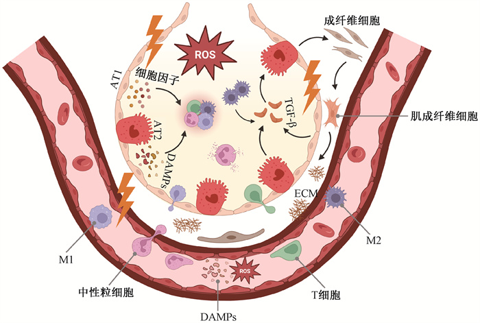
二、RILI中免疫细胞互作网络调控肺实质细胞的机制研究进展在RILI的发生、发展过程中,肺泡上皮细胞和肺血管内皮细胞作为辐射直接作用的关键靶细胞,通过释放DAMPs和细胞因子,激活免疫细胞表面受体,使其募集至损伤处;同时肺泡上皮细胞分泌IL-6、CXCL8等介质,进一步激活免疫细胞,被激活的免疫细胞释放ROS和促炎因子,加剧上皮损伤[50]。
在损伤后期,受损上皮细胞释放TGF-β等,诱导M2型巨噬细胞等免疫细胞分泌更多促纤维化因子,推动EMT进程,促进成纤维细胞活化增殖,导致细胞外基质(ECM)沉积,最终进展为不可逆纤维化[51](图 2)。
|
注:M1. M1型巨噬细胞;M2. M2型巨噬细胞;AT1. Ⅰ型肺泡上皮细胞;AT2. Ⅱ型肺泡上皮细胞;ROS. 活性氧;TGF-β. 转化生长因子β;ECM. 细胞外基质;DAMPs. 损伤相关分子模式 图 2 放射性肺损伤中免疫细胞与肺实质细胞交互调控肺纤维化进程示意图 Figure 2 Schematic diagram of the regulation of pulmonary fibrosis progression by the interaction between immune cells and lung parenchymal cells in radiation-induced lung injury |
1. 肺上皮细胞是RILI发生发展的核心环节:肺泡上皮细胞是肺组织的主要结构细胞,其损伤和功能障碍与放射性肺损伤的发生进展密切相关[52]。成熟的肺泡上皮细胞主要包括促进气体交换的Ⅰ型细胞(AT1)和分泌表面活性物质的Ⅱ型细胞(AT2)[53]。在RILI的早期炎症阶段,辐射诱导上皮细胞损伤释放DAMPs,启动先天免疫反应,招募中性粒细胞等免疫细胞浸润至损伤部位。受损的上皮细胞还分泌IL-6、TNF-α、IL-1β等促炎因子和CXCL8、CCL2等趋化因子,加剧局部炎症与组织水肿[50]。在正常情况下,AT2细胞可作为肺泡上皮的祖细胞进行增殖和分化以修复损伤,但持续的辐射损伤和炎症微环境可能抑制其修复能力,导致上皮再生障碍[54]。在放射性肺纤维化阶段,持续损伤的肺泡上皮细胞通过衰老相关分泌表型(SASP)释放TGF-β、PDGF等促纤维化因子,激活成纤维细胞,导致ECM的过度沉积和肺组织纤维化[51]。其中,TGF-β被电离辐射激活后可触发AT2细胞向间质细胞转化,上调间质标志物并抑制上皮标志物,进而产生大量ECM[55],是肺纤维化的核心细胞因子。
2. 肺血管内皮细胞通过多种途径参与RILI病理进程:肺血管内皮细胞是维持肺泡-毛细血管屏障功能的关键细胞[56]。肺血管内皮细胞受到电离辐射损伤后,不仅直接通过DNA损伤诱导细胞衰老,也间接通过释放DAMPs和SASP细胞因子促进巨噬细胞活化,导致血管通透性增加和血管完整性下降,进一步加剧了AT1和AT2细胞的损伤[57]。此外,血管内皮细胞获得促炎表型,上调ICAM-1、VCAM-1等黏附分子表达,促进中性粒细胞黏附和迁移,加重炎症反应[58]。受损血管的高通透性还导致纤维蛋白原外渗,经交联形成纤维蛋白并激活TGF-β,促进胶原沉积与纤维化发展[59]。
3. 肺成纤维细胞是放射性肺纤维化进展的主要推手:肺成纤维细胞是肺间质中的主要细胞类型,能够形成并维持富含ECM的多种结缔组织,然而在放射性肺损伤反复发生以及慢性疾病的情况下会引发病理性纤维化[60]。在损伤的初始阶段,成纤维细胞可被M1型巨噬细胞分泌的IL-1、IFN-γ等炎性细胞因子诱导分化为炎性成纤维细胞,而纤维化成纤维细胞随后被M2型巨噬细胞分泌的TGF-β1、PDGF等促纤维化细胞因子诱导[61]。其中,TGF-β/Smad信号通路在此过程中起核心作用,介导成纤维细胞向肌成纤维细胞转分化,导致ECM过度沉积、肺泡壁增厚及肺功能下降[62]。因此,成纤维细胞通过巨噬细胞及相关信号通路活化,是推动放射性肺纤维化进展的主要细胞。
三、靶向免疫细胞互作网络防治RILI的研究进展肺组织受辐照后多种免疫细胞募集活化,细胞间通过相互作用,共同参与放射性肺炎和纤维化的发展,因此靶向相关免疫细胞被认为是一种有效的治疗策略[63]。近年来,已有多项研究显示针对巨噬细胞、中性粒细胞、T细胞等免疫细胞的靶向干预药物可有效减轻RILI(表 1)。

|
表 1 靶向RILI微环境中不同免疫细胞的药物及作用机制 Table 1 Drugs targeting different immune cells in RILI and their mechanisms of action |
1. 靶向巨噬细胞极化状态干预RILI的策略:巨噬细胞的极化状态直接影响放射性肺损伤中的炎症反应和纤维化进程,因此靶向免疫细胞治疗RILI的相关研究目前主要集中在调控巨噬细胞极化方面。例如,吡非尼酮(PFD)通过抑制TGF-β1/Smad3通路调节巨噬细胞极化并改善辐射诱导的肺纤维化,减少胶原沉积和纤维化[64]。间充质干细胞(MSCs)作为炎症反应的调节剂,诱导M1型巨噬细胞向M2型巨噬细胞极化,缓解放射性肺炎[65]。氢疗法通过抑制NF-κB信号通路促进巨噬细胞从M1亚型极化到M2亚型,从而减轻对RILI中的炎症反应[66]。MCC950通过特异性抑制NLRP3炎性小体促进肺巨噬细胞向M2型极化,抑制炎症因子释放,从而缓解放射性肺炎[67]。岩藻多糖可以通过减少TIMP-1、CXCL1、MCP-1等炎性细胞因子表达以及巨噬细胞和中性粒细胞浸润来减轻辐射诱导的肺纤维化[68]。穿心莲内酯通过抑制AIM2炎性小体的激活,减少巨噬细胞焦亡,从而缓解TNF-α、IL-1β和IL-6等炎症因子的释放[69]。氯膦酸盐脂质体(LC)注射后被巨噬细胞吞噬和消化,在巨噬细胞内释放氯膦酸钠,诱导巨噬细胞凋亡,减轻辐射诱导的肺损伤[70]。上述相关药物中,除PFD已进展到临床试验阶段[71],其余大部分药物基于动物模型初步探索了其可能的作用机制,未来仍需进一步加强基础研究和临床试验,以推动其在RILI治疗中的广泛应用。
2. 其他靶向免疫细胞防治RILI的探索:近年来,靶向其他免疫细胞的RILI防治策略也取得了一定进展。例如,中性粒细胞在RILI中主要参与早期炎症反应,其过度活化会导致组织损伤。西维来司他钠通过抑制中性粒细胞释放中性粒细胞弹性蛋白酶来抑制辐射诱导的肺损伤,因此在RILI早期阶段给药被认为可能更加有效[72]。T细胞亚群在放射性肺损伤时失衡,调节T细胞亚群平衡被认为可减轻炎症反应。例如白藜芦醇苷通过抑制TGF-β1/Smad3信号通路EMT,缓解辐射诱导的Th1/Th2失衡,从而减轻辐射诱导的肺损伤[73]。
目前靶向中性粒细胞及T细胞等免疫细胞防治RILI的策略仍处于初步探索阶段。例如,西维来司他钠抑制中性粒细胞过度活化、白藜芦醇苷调节T细胞亚群平衡等研究,大多基于动物实验验证其潜在机制与疗效,尚未进入临床转化阶段。未来需进一步明确其作用途径、用药时机及长期安全性,推动更多针对免疫微环境多细胞协同调控的干预策略研发,为RILI的防治提供新方向。
四、总结与展望放射性肺损伤是胸部肿瘤放疗后常见的并发症,多种免疫细胞通过释放细胞因子、趋化因子和活性氧等介质,推动炎症反应和纤维化进程。尽管近年来对免疫细胞在RILI中的作用机制有了更深入的理解,但仍有许多问题亟待解决。例如多种免疫细胞参与RILI的具体机制缺乏深入研究,肿瘤微环境中的免疫细胞分布对RILI的影响尚不明确,放射治疗与免疫检查点抑制剂(如PD-1/PD-L1抑制剂)联合使用在某些情况下可增强抗肿瘤效果,但同时也可能增加放射性肺损伤的风险。未来还需深入了解各类免疫细胞中互作网络的动态变化并结合肿瘤免疫微环境对RILI免疫细胞的作用机制进一步探索。
同时,针对免疫细胞的靶向治疗已表现出巨大潜力。例如,相关研究发现食管癌患者同步放化疗时联合PFD治疗,既能有效降低RILI发生率、改善患者肺功能,又有助于提高肿瘤控制效果、延长患者生存期[71]。此外,利用单细胞测序等新技术,能够精确解析免疫细胞亚群的功能和分子特征,从而发现新的治疗靶点和生物标志物,推动放射性肺损伤的精准治疗[74]。综上,深入理解免疫细胞在RILI中的互作机制,有望为临床上开发靶向治疗策略、改善患者预后和生活质量提供新思路,从而推进放疗在肿瘤治疗中的应用。
利益冲突 无
作者贡献声明 曹诗雯负责文献搜集整理和论文撰写;焦旸指导论文修改
| [1] |
Giuranno L, Ient J, De Ruysscher D, et al. Radiation-induced lung injury (RILI)[J]. Front Oncol, 2019, 9: 877. DOI:10.3389/fonc.2019.00877 |
| [2] |
Deng G, Liang N, Xie J, et al. Pulmonary toxicity generated from radiotherapeutic treatment of thoracic malignancies[J]. Oncol Lett, 2017, 14(1): 501-511. DOI:10.3892/ol.2017.6268 |
| [3] |
姜秀政, 华玥祺, 田振桃, 等. 放射性肺损伤的研究进展[J]. 医学理论与实践, 2021, 34(10): 1651-1653. Jiang XZ, Hua YQ, Tian ZT, et al. Research progress of radiation-induced lung injury[J]. J Med Theo Prac, 2021, 34(10): 1651-1653. DOI:10.19381/j.issn.1001-7585.2021.10.010 |
| [4] |
Hanania AN, Mainwaring W, Ghebre YT, et al. Radiation-induced lung injury: Assessment and management[J]. Chest, 2019, 156(1): 150-162. DOI:10.1016/j.chest.2019.03.033 |
| [5] |
Feldman N, Rotter-Maskowitz A, Okun E. DAMPs as mediators of sterile inflammation in aging-related pathologies[J]. Ageing Res Rev, 2015, 24(Pt A): 29-39. DOI:10.1016/j.arr.2015.01.003 |
| [6] |
Ayass MA, Tripathi T, Zhu K, et al. T helper (Th) cell profiles and cytokines/chemokines in characterization, treatment, and monitoring of autoimmune diseases[J]. Methods, 2023, 220: 115-125. DOI:10.1016/j.ymeth.2023.11.003 |
| [7] |
Vasse GF, Nizamoglu M, Heijink IH, et al. Macrophage-stroma interactions in fibrosis: Biochemical, biophysical, and cellular perspectives[J]. J Pathol, 2021, 254(4): 344-357. DOI:10.1002/path.5632 |
| [8] |
Nathan C. Neutrophils and immunity: Challenges and opportunities[J]. Nat Rev Immunol, 2006, 6(3): 173-182. DOI:10.1038/nri1785 |
| [9] |
Aurora AB, Porrello ER, Tan W, et al. Macrophages are required for neonatal heart regeneration[J]. J Clin Invest, 2014, 124(3): 1382-1392. DOI:10.1172/JCI72181 |
| [10] |
Weaver CT, Elson CO, Fouser LA, et al. The Th17 pathway and inflammatory diseases of the intestines, lungs, and skin[J]. Annu Rev Pathol, 2013, 8: 477-512. DOI:10.1146/annurev-pathol-011110-130318 |
| [11] |
Braciale TJ, Sun J, Kim TS. Regulating the adaptive immune response to respiratory virus infection[J]. Nat Rev Immunol, 2012, 12(4): 295-305. DOI:10.1038/nri3166 |
| [12] |
Zhang Z, Zhou J, Verma V, et al. Crossed pathways for radiation-induced and immunotherapy-related lung injury[J]. Front Immunol, 2021, 12: 774807. DOI:10.3389/fimmu.2021.774807 |
| [13] |
Wang L, Zhao W, Ning X, et al. Effect of X-ray irradiation combined with PD-1 inhibitor treatment on lung tissue injury in mice[J]. Int Immunopharmacol, 2023, 123: 110775. DOI:10.1016/j.intimp.2023.110775 |
| [14] |
Li J, Zeng Z, Wu Q, et al. Immunological modulation of the Th1/Th2 shift by ionizing radiation in tumors (Review)[J]. Int J Oncol, 2021, 59(1). DOI:10.3892/ijo.2021.5230 |
| [15] |
Paun A, Bergeron M, Haston CK. The Th1/Th17 balance dictates the fibrosis response in murine radiation-induced lung disease[J]. Sci Rep, 2017, 7(1): 11586. DOI:10.1038/s41598-017-11656-5 |
| [16] |
Chen J, Wang Y, Mei Z, et al. Radiation-induced lung fibrosis in a tumor-bearing mouse model is associated with enhanced Type-2 immunity[J]. J Radiat Res, 2016, 57(2): 133-141. DOI:10.1093/jrr/rrv077 |
| [17] |
Park H, Li Z, Yang XO, et al. A distinct lineage of CD4 T cells regulates tissue inflammation by producing interleukin 17[J]. Nat Immunol, 2005, 6(11): 1133-1141. DOI:10.1038/ni1261 |
| [18] |
Sun L, Su Y, Jiao A, et al. T cells in health and disease[J]. Signal Transduct Target Ther, 2023, 8(1): 235. DOI:10.1038/s41392-023-01471-y |
| [19] |
Yang X, Walton W, Cook DN, et al. The chemokine, CCL3, and its receptor, CCR1, mediate thoracic radiation-induced pulmonary fibrosis[J]. Am J Respir Cell Mol Biol, 2011, 45(1): 127-135. DOI:10.1165/rcmb.2010-0265OC |
| [20] |
Carbone F, Nencioni A, Mach F, et al. Pathophysiological role of neutrophils in acute myocardial infarction[J]. Thromb Haemost, 2013, 110(3): 501-514. DOI:10.1160/TH13-03-0211 |
| [21] |
Collison LW, Workman CJ, Kuo TT, et al. The inhibitory cytokine IL-35 contributes to regulatory T-cell function[J]. Nature, 2007, 450(7169): 566-569. DOI:10.1038/nature06306 |
| [22] |
Joetham A, Takeda K, Taube C, et al. Naturally occurring lung CD4(+)CD25(+) T cell regulation of airway allergic responses depends on IL-10 induction of TGF-beta[J]. J Immunol, 2007, 178(3): 1433-1442. DOI:10.4049/jimmunol.178.3.1433 |
| [23] |
Tan W, Zhang C, Liu J, et al. Regulatory T-cells promote pulmonary repair by modulating T helper cell immune responses in lipopolysaccharide-induced acute respiratory distress syndrome[J]. Immunology, 2019, 157(2): 151-162. DOI:10.1111/imm.13060 |
| [24] |
Xiong S, Guo R, Yang Z, et al. Treg depletion attenuates irradiation-induced pulmonary fibrosis by reducing fibrocyte accumulation, inducing Th17 response, and shifting IFN-γ, IL-12/IL-4, IL-5 balance[J]. Immunobiology, 2015, 220(11): 1284-1291. DOI:10.1016/j.imbio.2015.07.001 |
| [25] |
Wirsdörfer F, de Leve S, Cappuccini F, et al. Extracellular adenosine production by ecto-5'-Nucleotidase (CD73) enhances radiation-induced lung fibrosis[J]. Cancer Res, 2016, 76(10): 3045-3056. DOI:10.1158/0008-5472.CAN-15-2310 |
| [26] |
Xiong S, Pan X, Xu L, et al. Regulatory T cells promote β-catenin--mediated epithelium-to-mesenchyme transition during radiation-induced pulmonary fibrosis[J]. Int J Radiat Oncol Biol Phys, 2015, 93(2): 425-435. DOI:10.1016/j.ijrobp.2015.05.043 |
| [27] |
House IG, Savas P, Lai J, et al. Macrophage-derived CXCL9 and CXCL10 are required for antitumor immune responses following immune checkpoint blockade[J]. Clin Cancer Res, 2020, 26(2): 487-504. DOI:10.1158/1078-0432.CCR-19-1868 |
| [28] |
Yahyapour R, Shabeeb D, Cheki M, et al. Radiation protection and mitigation by natural antioxidants and flavonoids: implications to radiotherapy and radiation disasters[J]. Curr Mol Pharmacol, 2018, 11(4): 285-304. DOI:10.2174/1874467211666180619125653 |
| [29] |
Zhang J, Liu X, Wan C, et al. NLRP3 inflammasome mediates M1 macrophage polarization and IL-1β production in inflammatory root resorption[J]. J Clin Periodontol, 2020, 47(4): 451-460. DOI:10.1111/jcpe.13258 |
| [30] |
Ni J, Guo T, Zhou Y, et al. STING signaling activation modulates macrophage polarization via CCL2 in radiation-induced lung injury[J]. J Transl Med, 2023, 21(1): 590. DOI:10.1186/s12967-023-04446-3 |
| [31] |
Liu X, Zeng L, Zhou Y, et al. P21 facilitates macrophage chemotaxis by promoting CCL7 in the lung epithelial cell lines treated with radiation and bleomycin[J]. J Transl Med, 2023, 21(1): 314. DOI:10.1186/s12967-023-04177-5 |
| [32] |
Van Dyken SJ, Locksley RM. Interleukin-4- and interleukin-13-mediated alternatively activated macrophages: Roles in homeostasis and disease[J]. Annu Rev Immunol, 2013, 31: 317-343. DOI:10.1146/annurev-immunol-032712-095906 |
| [33] |
Xu Q, Choksi S, Qu J, et al. NADPH oxidases are essential for macrophage differentiation[J]. J Biol Chem, 2016, 291(38): 20030-20041. DOI:10.1074/jbc.M116.731216 |
| [34] |
Park HR, Jo SK, Jung U. Ionizing radiation promotes epithelial-to-mesenchymal transition in lung epithelial cells by TGF-β-producing M2 macrophages[J]. In Vivo, 2019, 33(6): 1773-1784. DOI:10.21873/invivo.11668 |
| [35] |
Bhandari R, Ball MS, Martyanov V, et al. Profibrotic activation of human macrophages in systemic sclerosis[J]. Arthritis Rheumatol, 2020, 72(7): 1160-1169. DOI:10.1002/art.41243 |
| [36] |
Kolaczkowska E, Kubes P. Neutrophil recruitment and function in health and inflammation[J]. Nat Rev Immunol, 2013, 13(3): 159-175. DOI:10.1038/nri3399 |
| [37] |
Gupta AK, Joshi MB, Philippova M, et al. Activated endothelial cells induce neutrophil extracellular traps and are susceptible to NETosis-mediated cell death[J]. FEBS Lett, 2010, 584(14): 3193-3197. DOI:10.1016/j.febslet.2010.06.006 |
| [38] |
Liew PX, Kubes P. The neutrophil's role during health and disease[J]. Physiol Rev, 2019, 99(2): 1223-1248. DOI:10.1152/physrev.00012.2018 |
| [39] |
Siwicki M, Kubes P. Neutrophils in host defense, healing, and hypersensitivity: Dynamic cells within a dynamic host[J]. J Allergy Clin Immunol, 2023, 151(3): 634-655. DOI:10.1016/j.jaci.2022.12.004 |
| [40] |
Li Y, Zou L, Chu L, et al. Identification and integrated analysis of circRNA and miRNA of radiation-induced lung injury in a mouse model[J]. J Inflamm Res, 2021, 14: 4421-4431. DOI:10.2147/JIR.S322736 |
| [41] |
Mauri C, Gray D, Mushtaq N, et al. Prevention of arthritis by interleukin 10-producing B cells[J]. J Exp Med, 2003, 197(4): 489-501. DOI:10.1084/jem.20021293 |
| [42] |
Liu B, Wang Y, Han G, et al. Tolerogenic dendritic cells in radiation-induced lung injury[J]. Front Immunol, 2023, 14: 1323676. DOI:10.3389/fimmu.2023.1323676 |
| [43] |
Vivier E, Tomasello E, Baratin M, et al. Functions of natural killer cells[J]. Nat Immunol, 2008, 9(5): 503-510. DOI:10.1038/ni1582 |
| [44] |
Wang R, Ma X, Zhang X, et al. Autophagy-mediated NKG2D internalization impairs NK cell function and exacerbates radiation pneumonitis[J]. Front Immunol, 2023, 14: 1250920. DOI:10.3389/fimmu.2023.1250920 |
| [45] |
Bocchino M, Zanotta S, Capitelli L, et al. Dendritic cells are the intriguing players in the puzzle of idiopathic pulmonary fibrosis pathogenesis[J]. Front Immunol, 2021, 12: 664109. DOI:10.3389/fimmu.2021.664109 |
| [46] |
Ferris ST, Durai V, Wu R, et al. cDC1 prime and are licensed by CD4+ T cells to induce anti-tumour immunity[J]. Nature, 2020, 584(7822): 624-629. DOI:10.1038/s41586-020-2611-3 |
| [47] |
Liu B, Wang Y, Ma L, et al. CCL22 induces the polarization of immature dendritic cells into tolerogenic dendritic cells in radiation-induced lung injury through the CCR4-Dectin2-PLC-γ2-NFATC2-Nr4a2-PD-L1 signaling pathway[J]. J Immunol, 2024, 213(3): 268-282. DOI:10.4049/jimmunol.2300718 |
| [48] |
Mascanfroni ID, Yeste A, Vieira SM, et al. IL-27 acts on DCs to suppress the T cell response and autoimmunity by inducing expression of the immunoregulatory molecule CD39[J]. Nat Immunol, 2013, 14(10): 1054-1063. DOI:10.1038/ni.2695 |
| [49] |
Volchenkov R, Karlsen M, Jonsson R, et al. Type 1 regulatory T cells and regulatory B cells induced by tolerogenic dendritic cells[J]. Scand J Immunol, 2013, 77(4): 246-254. DOI:10.1111/sji.12039 |
| [50] |
Fleckenstein K, Gauter-Fleckenstein B, Jackson IL, et al. Using biological markers to predict risk of radiation injury[J]. Semin Radiat Oncol, 2007, 17(2): 89-98. DOI:10.1016/j.semradonc.2006.11.004 |
| [51] |
Coppé J, Desprez P, Krtolica A, et al. The senescence-associated secretory phenotype: The dark side of tumor suppression[J]. Annu Rev Pathol, 2010, 5: 99-118. DOI:10.1146/annurev-pathol-121808-102144 |
| [52] |
Satyamitra MM, DiCarlo AL, Taliaferro L. Understanding the pathophysiology and challenges of development of medical countermeasures for radiation-induced vascular/endothelial cell injuries: report of a NIAID workshop, August 20, 2015[J]. Radiat Res, 2016, 186(2): 99-111. DOI:10.1667/RR14436.1 |
| [53] |
Jain R, Barkauskas CE, Takeda N, et al. Plasticity of Hopx+ type Ⅰ alveolar cells to regenerate type Ⅱ cells in the lung[J]. Nat Commun, 2015, 6: 6727. DOI:10.1038/ncomms7727 |
| [54] |
Yang M, Fan Q, Hei TK, et al. Single-cell transcriptome analysis of radiation pneumonitis mice[J]. Antioxidants (Basel), 2022, 11(8): 1457. DOI:10.3390/antiox11081457 |
| [55] |
Sisto M, Ribatti D, Lisi S. Organ fibrosis and autoimmunity: The role of inflammation in TGF β-dependent EMT[J]. Biomolecules, 2021, 11(2): 310. DOI:10.3390/biom11020310 |
| [56] |
Aird WC. Endothelial cell heterogeneity[J]. Cold Spring Harb Perspect Med, 2012, 2(1): a006429. DOI:10.1101/cshperspect.a006429 |
| [57] |
Fleckenstein K, Zgonjanin L, Chen L, et al. Temporal onset of hypoxia and oxidative stress after pulmonary irradiation[J]. Int J Radiat Oncol Biol Phys, 2007, 68(1): 196-204. DOI:10.1016/j.ijrobp.2006.12.056 |
| [58] |
Soroush F, Tang Y, Zaidi HM, et al. PKCδ inhibition as a novel medical countermeasure for radiation-induced vascular damage[J]. FASEB J, 2018, fj201701099. DOI:10.1096/fj.201701099 |
| [59] |
Laurens N, Koolwijk P, de Maat MPM. Fibrin structure and wound healing[J]. J Thromb Haemost, 2006, 4(5): 932-939. DOI:10.1111/j.1538-7836.2006.01861.x |
| [60] |
Henderson NC, Rieder F, Wynn TA. Fibrosis: From mechanisms to medicines[J]. Nature, 2020, 587(7835): 555-566. DOI:10.1038/s41586-020-2938-9 |
| [61] |
Tsukui T, Wolters PJ, Sheppard D. Alveolar fibroblast lineage orchestrates lung inflammation and fibrosis[J]. Nature, 2024, 631(8021): 627-634. DOI:10.1038/s41586-024-07660-1 |
| [62] |
Zhang YE. Non-smad signaling pathways of the TGF-β family[J]. Cold Spring Harb Perspect Biol, 2017, 9(2): a022129. DOI:10.1101/cshperspect.a022129 |
| [63] |
王丽兴, 勾文峰, 许飞飞, 等. 放射性肺损伤免疫微环境的变化及治疗策略[J]. 沈阳药科大学学报, 2025, 42(1): 1-9. Wang LX, Gou WF, Xu FF, et al. Changes in the immune microenvironment of radiation-induced lung injury and therapeutic strategies[J]. J Shenyang Pharma Univ, 2025, 42(1): 1-9. DOI:10.14066/j.cnki.cn21-1349/r.2024.0345 |
| [64] |
Ying H, Fang M, Hang QQ, et al. Pirfenidone modulates macrophage polarization and ameliorates radiation-induced lung fibrosis by inhibiting the TGF-β1/Smad3 pathway[J]. J Cell Mol Med, 2021, 25(18): 8662-8675. DOI:10.1111/jcmm.16821 |
| [65] |
荆云涛, 安广洲, 来盼盼, 等. 脐带间充质干细胞对放射性肺损伤的防治作用[J]. 空军军医大学学报, 2023, 44(3): 203-209. Jing YT, An GZ, Lai PP, et al. Preventive and therapeutic effects of umbilical cord mesenchymal stem cells on radiation-induced lung injury[J]. J Air Force Med Univ, 2023, 44(3): 203-209. DOI:10.13276/j.issn.2097-1656.2023.03.003 |
| [66] |
Gao X, Niu S, Li L, et al. Hydrogen therapy promotes macrophage polarization to the M2 subtype in radiation lung injury by inhibiting the NF-κB signalling pathway[J]. Heliyon, 2024, 10(10): e30902. DOI:10.1016/j.heliyon.2024.e30902 |
| [67] |
王贞, 路素丽, 何贵成, 等. MCC950调控巨噬细胞极化改善放射性肺损伤[J]. 辐射研究与辐射工艺学报, 2024, 42(5): 56-64. Wang Z, Lu SL, He GC, et al. MCC950 ameliorates radiation induced lung injury through modulating macrophage polarization[J]. J Radiat Res Radiat Process, 2024, 42(5): 56-64. DOI:10.11889/j.1000-3436.2024-0026 |
| [68] |
Yu HH, Chengchuan Ko E, Chang CL, et al. Fucoidan inhibits radiation-induced pneumonitis and lung fibrosis by reducing inflammatory cytokine expression in lung tissues[J]. Mar Drugs, 2018, 16(10): 392. DOI:10.3390/md16100392 |
| [69] |
Gao J, Peng S, Shan X, et al. Inhibition of AIM2 inflammasome-mediated pyroptosis by andrographolide contributes to amelioration of radiation-induced lung inflammation and fibrosis[J]. Cell Death Dis, 2019, 10(12): 957. DOI:10.1038/s41419-019-2195-8 |
| [70] |
Song G, Cai F, Liu L, et al. Liposomal sodium clodronate mitigates radiation-induced lung injury through macrophage depletion[J]. Transl Oncol, 2024, 47: 102029. DOI:10.1016/j.tranon.2024.102029 |
| [71] |
邱国钦, 陈肖珏, 许英艺, 等. 吡非尼酮防治食管癌放射性肺损伤的临床观察[J]. 肿瘤防治研究, 2025, 52(3): 217-224. Qiu GQ, Chen XJ, Xu YY, et al. Clinical observation of pirfenidone in prevention and treatment of radiation-induced lung injury in esophageal cancer[J]. Cancer Res Prev Treat, 2025, 52(3): 217-224. DOI:10.3971/j.issn.1000-8578.2025.24.0592 |
| [72] |
Yoshikawa N, Inomata T, Okada Y, et al. Sivelestat sodium hydrate reduces radiation-induced lung injury in mice by inhibiting neutrophil elastase[J]. Mol Med Rep, 2013, 7(4): 1091-1095. DOI:10.3892/mmr.2013.1318 |
| [73] |
Cao K, Lei X, Liu H, et al. Polydatin alleviated radiation-induced lung injury through activation of Sirt3 and inhibition of epithelial-mesenchymal transition[J]. J Cell Mol Med, 2017, 21(12): 3264-3276. DOI:10.1111/jcmm.13230 |
| [74] |
Wu F, Zhang Z, Wang M, et al. Cellular atlas of senescent lineages in radiation- or immunotherapy-induced lung injury by single-cell RNA-sequencing analysis[J]. Int J Radiat Oncol Biol Phys, 2023, 116(5): 1175-1189. DOI:10.1016/j.ijrobp.2023.02.005 |