 2026, Vol. 46


2. 飞利浦医疗临床科研部, 成都 610041;
3. 福禄克测试仪器(上海)有限公司, 上海 200335;
4. 上海西门子医疗器械有限公司, 上海 201318
2. Philips Healthcare Clinical Research Department, Chengdu 610041, China;
3. Fluke Test Instruments (Shanghai) Co., Ltd., Shanghai 200335, China;
4. Siemens Shanghai Medical Equipment Ltd., Shanghai 201318, China
CT检查是公众医源性电离辐射的主要来源,其潜在致癌风险备受关注[1-5]。业界持续优化扫描参数及重建算法,力求降低辐射剂量[6-10]。铅屏蔽作为防护手段之一,可减少散射辐射,对敏感器官形成定向保护[11-12]。而铅屏蔽效能有显著位置依赖性。若屏蔽体靠近主射线束,其高衰减特性可被自动曝光控制系统(automatic exposure control,AEC)误判为高密度组织,触发管电流补偿性增加,升高受检者剂量[13-16]。现有共识建议铅屏蔽须置于扫描野外,且距主射线束边界≥5 cm[17-18]。但有研究提示,即使距离达7.5 cm,大螺距扫描时仍可能干扰曝光控制系统[19]。铅屏蔽放置位置对AEC影响及对敏感器官剂量降低效果缺乏基于不同探测器宽度的系统评估。本研究通过多机型体模实验,探究铅屏蔽放置位置对6种不同探测器宽度机型AEC的干扰情况,并测量性腺模拟点剂量变化,旨在建立基于探测器的个体化防护模型,为精准辐射防护提供实证依据。
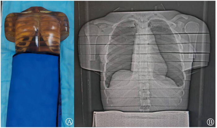
材料与方法1. 一般材料:采用成都方拓仿真技术有限责任公司的中国人辐照仿真人体模型,如图 1所示。本研究所用铅单巾铅当量为0.5 mmPb。屏蔽方式均采用360°包裹式屏蔽。
|
图 1 仿真人体模型(A)与胸部扫描时仿真人体模型的定位图像(B) Figure 1 Anthropomorphic phantom (A) and its scout image during chest scanning (B) |
2.CT扫描:使用德国西门子16排Scope、32排Go Up、64排Flash、96排Force、光子计数Alpha等不同探测器宽度的机型对体模进行CT胸部扫描。扫描方向自头向足,扫描范围肺尖至肺底,总长度为251 mm。各设备扫描参数列于表 1。每台设备均使用Care Dose 4D进行自动mA调制。根据铅单巾上缘与扫描野下缘的距离不同分为4组:0(2组)、2(3组)、4(4组)和6 cm(5组),无防护(1组)作为对照组。每组行10次扫描。使用容积CT剂量指数(volume computed tomography dose index,CTDIvol)和剂量长度乘积(dose length product,DLP)评估每台设备胸部扫描区域整体辐射剂量。采用Raysafe X2空气电离室CT探头(上海Fluke Biomedical公司,RaySafe X2,CT Sensor,简称RX2,量程为10 μGy~999 Gy) 测量体模的女性生殖器平面表面和中心、男性生殖器表面吸收剂量,将探头紧贴男性生殖器(睾丸)及盆腔测量其受照射情况,将探头水平置于在脐与髂前上棘连线的中点与耻骨联合中点间作连线中点层面,模拟女性生殖器(卵巢)受照射情况。使用德国西门子原厂软件重建扫描数据,重建层厚为5 mm,使用西门子自带图像浏览软件记录每层的管电流,计算变化量,反映铅单巾对AEC的影响。

|
表 1 各设备扫描参数 Table 1 Technical scanning parameters for each CT system |
3.统计学处理:使用SPSS 27.0软件进行统计学分析。计量资料符合正态分布,用 x±s表示。数据进行单因素方差分析,组间进行两两比较,方差齐使用Tukey HSD检验,不齐使用Dunnett T3检验。P<0.05为差异具有统计学意义。计算各组每层管电流均值
1.铅单巾放置于不同防护位置的胸部CTDIvol和DLP比较:铅单巾放置于不同防护位置的CTDvol和DLP列于表 2,3。铅单巾距扫描野的距离增加显著影响扫描区域整体辐射剂量指标,CTDIvol与DLP呈现同步变化趋势。Scope (探测器宽度19.2 mm)、光子薄层(探测器宽度19.2 mm)在2 cm距离时,CTDIvol差异<1%。32排Go up(探测器宽度22.4 mm)在2 cm时CTDIvol较无防护升高1.3%,DLP同步升高1.1%,需增至4 cm距离可使CTDIvol与DLP差异<1%。64排Flash(38.4 mm)在4 cm时CTDIvol显著升高4.9%,DLP升高4.7%;至6 cm时CTDIvol差异缩小至0.88%,DLP同步缩小至0.97%。96排Force(57.6 mm)在6 cm时CTDIvol差异为2.1%,DLP同步为2.2%。光子常规(57.6 mm)在6 cm时CTDIvol、DLP与无防护差异<1%。
|
|
表 2 铅单巾放置于不同防护位置的胸部扫描区域整体CTDIvol比较(mGy,x±s) Table 2 Comparison of overall chest CTDIvol in the scanning area with lead aprons placed at different protective positions (mGy, x±s) |
|
|
表 3 铅单巾放置于不同防护位置的胸部扫描区域整体DLP比较(mGy·cm,x±s) Table 3 Comparison of overall chest DLP in the scanning area with lead aprons placed at different protective positions(mGy·cm, x±s) |
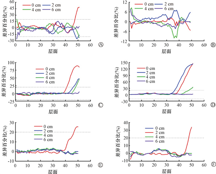
2.铅单巾放置于不同防护位置各个层面mA分布:西门子16排Scope、64排Flash、96排Force重建层数为51层。32排Go Up与光子计数Alpha为50层。所有机型的管电流均值差异如图 2所示。在近距离防护(0 cm)时,Scope、Flash、Force、光子常规、光子薄层出现管电流异常升高(>20%差异)。Scope机型(19.2 mm)在0 cm防护时管电流异常升高层面为4(长度2 mm),与探测器宽度相当,距离增至2 cm异常层面消失。Flash(38.4 mm)机型在0 cm时异常层面宽50 mm,至6 cm距离时完全恢复正常。Force (57.6 mm)机型在0 cm时异常层面宽75 mm,6 cm时差异消失。光子常规(57.6 mm)扫描在0 cm时异常层面宽20 mm,2 cm时恢复正常;光子薄层在0 cm时异常层面宽15 mm存在差异,2 cm时恢复。Go up管电流差异未超过20%。
|
图 2 不同型号CT中铅单巾放置于不同防护位置的各层面mA差异比较 A. Scope;B. Go up;C. Flash;D. Force;E. 光子常规;F. 光子薄层 Figure 2 Tube current (mA) variations with lead aprons placed at different protective in different CT scaner A. Scope; B. Go up; C. Flash; D. Force; E. Photon-counting CT 144-slice scanning mode; F. Photon-counting CT 96-slice scanning mode |
3.性腺的吸收剂量:性腺的吸收剂量列于表 4。无防护时女性性腺剂量在Scope机型仅占胸部扫描剂量0.99%,Go up机型为1.28%,Flash机型为0.88%,Force机型为1.03%,光子常规为0.85%,光子薄层为0.73%。无防护时各机型的男性性腺剂量测量值分别为:Scope为(0.044 5±0.000 5)mGy;Go up为(0.054 2±0.000 9)mGy;Flash为(0.029 8±0.000 8)mGy;Force为(0.028 7±0.000 2)mGy;光子常规为(0.036 7±0.000 3)mGy;光子薄层为(0.032 0±0.000 2)mGy。无防护时男性性腺剂量Scope机型上占胸部扫描剂量0.63%,Go up机型为0.97%,Flash机型为0.67%,Force机型为0.85%,光子常规为0.86%,光子薄层为0.71%。女性卵巢剂量在防护后降低。Scope、Go up、Force、光子常规、光子薄层能在铅屏蔽0 cm获得最大防护收益,最大可达76.9%(Go up)。不同防护距离下,性腺的吸收剂量均值最大差异为0.008 5 mGy。所有防护距离下男性睾丸位置均未检出可测量剂量。
|
|
表 4 铅单巾放置于不同防护位置的女性性腺吸收剂量(RX 2测量值)对比(mGy, x±s) Table 4 Female gonadal radiation dose comparison with lead aprons placed at different protective (mGy, x±s) |
讨论
本研究系统评估了铅单巾放置位置对不同CT机型胸部扫描中散射至性腺区域剂量的影响,以及其对扫描剂量指标(CTDIvol, DLP)及管电流调制(mA)分布的影响。
研究发现,当铅单巾放置过近,其边缘进入或靠近当前扫描的探测器Z轴有效覆盖范围时,会部分阻挡入射X射线束。Care Dose 4D等自动管电流调制系统误判该区域需要更高剂量以达到预设图像质量,会显著提升相应扫描位置(通常是扫描野足侧末端层面)的管电流(造成>20%差异)。这种现象是自动管电流调制系统遇到非患者解剖结构(如铅单巾)干扰时的直接表现[13-16],仅有当铅单巾需放置在足够远处,才能避免不必要的mA补偿,降低胸部吸收剂量。铅单巾放置位置是否影响AEC与探测器宽度息息相关。较窄的探测器(如Scope 19.2 mm, Go Up 22.4 mm, 光子薄层19.2 mm)在略大于其探测器宽度的防护距离下可使剂量指标(CTDIvol,DLP)接近无防护水平(差异<2%),且管电流异常升高的范围消失。相反,较宽的探测器(如Flash 38.4 mm, Force 57.6 mm, 光子常规57.6 mm)则需要更远的防护位置才能使胸部剂量指标差异降至<2%(最小值为0.002 mGy,最大为0.072 mGy)。有研究报道,安装时间不同的两台Force对于胸部前后径平均值相近的患者(22.66 cm vs 22.83 cm)胸部扫描剂量差异达到0.25 mGy[21]。合理铅单巾位置引起的差异已明显小于其他因素引起的剂量差异。本研究发现设置铅单巾后,不同机型在不同扫描层面mA所受影响呈现异质性。这种差异可能与各机型所采用的图像重建算法及曝光控制中的自动管电流调制机制有关。
无防护时所有机型的男性、女性性腺模拟位置的吸收剂量均未超过扫描区域整体的剂量指标的1.5%,大部分机型不足1%。针对模拟女性性腺,若铅单巾放置位置不当,虽可降低性腺剂量,但会引起扫描区域整体剂量指标的显著上升。这表明,铅单巾的设置位置应依据受检者所需保护的具体器官进行针对性选择,以实现防护效果与扫描剂量之间的最佳平衡。不同防护位置下,性腺的吸收剂量差异小,最大差异(Go Up:0与4 cm)不足0.009 mGy,此时扫描区域整体CTDIvol差异达到0.11 mGy。对于女性患者,设置铅单巾可以减少性腺55%~76%的吸收剂量,设置后不同位置的吸收差异不大,而扫描区域整体的剂量指标差异较大,此时可以考虑铅单巾放置不同位置对扫描区域整体的剂量指标的干扰。所有机型在保证扫描区域整体的剂量指标增幅≤2.1%,性腺吸收剂量降幅仍可达55.6%~70.3%(如Scope-2 cm降70.3%,Force-6 cm降59.1%)。所有防护距离下紧贴体表睾丸位置的RX 2均未测到可记录剂量。这可能因为睾丸位于扫描野外体表,散射辐射本底本身已很低,且铅屏蔽阻挡效果更直接。以往研究得到相似的结果[12]。这一结果提示,在常规胸部CT中,对于男性患者,设置铅屏蔽后降低性腺吸收剂量,降低的值与铅单巾设置位置相关性不强,可重点考虑扫描区域整体的剂量指标的影响。本研究证实,在胸部CT扫描中,根据不同探测器宽度确定铅单巾合理放置位置,可降低女性性腺区域的辐射剂量且对计划扫描区域的剂量(CTDIvol和DLP)影响较小。
本研究尚存在一定的局限性。首先,实验基于标准体模,可能与真实人体对剂量分布的准确性存在一定的影响。其次,睾丸敏感器官的测量结果为阴性,该结果可能需使用更灵敏的剂量计或在更贴近真实组织深度的条件下进一步验证。第三,所有数据均来源于西门子CT设备及其Care Dose 4D自动曝光控制系统,结论外推至其他设备需谨慎。第四,本研究采用Raysafe X2空气电离室CT探头进行测量,而未使用热释光剂量计,这可能对深部或小视野区域的剂量评估精度造成一定影响,课题组未来将采用热释光剂量计在患者真实场景上进行测量。最后,本研究未探讨管电压、螺距等其他扫描参数对防护效果的影响;实验仅针对特定敏感器官的散射剂量进行测量,未能全面评估非扫描区域的辐射剂量空间分布及其潜在健康风险,有待后续研究进一步完善。
利益冲突 无
作者贡献声明 文大光负责实验设计、数据处理、数据分析、论文撰写和修改;宋婷妮、负责实验设计、论文修改;张晓咏、彭晶参与论文撰写与修改;宋建安、叶硕奇、田毅参与技术指导与论文修改;李真林、夏春潮制定研究方向、负责实验设计、指导论文修改
| [1] |
Smith-Bindman R, Chu PW, Azman Firdaus H, et al. Projected lifetime cancer risks from current computed tomography imaging[J]. JAMA Intern Med, 2025, 185(6): 710-719. DOI:10.1001/jamainternmed.2025.0505 |
| [2] |
Bosch de Basea M, Moriña D, Figuerola J, et al. Subtle excess in lifetime cancer risk related to CT scanning in Spanish young people[J]. Environ Int, 2018, 120: 1-10. DOI:10.1016/j.envint.2018.07.020 |
| [3] |
Lee KH, Lee S, Park JH, et al. Risk of hematologic malignant neoplasms from abdominopelvic computed tomographic radiation in patients who underwent appendectomy[J]. JAMA Surg, 2021, 156(4): 343-351. DOI:10.1001/jamasurg.2020.6357 |
| [4] |
Hauptmann M, Byrnes G, Cardis E, et al. Brain cancer after radiation exposure from CT examinations of children and young adults: results from the EPI-CT cohort study[J]. Lancet Oncol, 2023, 24(1): 45-53. DOI:10.1016/S1470-2045(22)00655-6 |
| [5] |
邓小琴, 喻崇容, 彭燕, 等. 胸部CT扫描技术与辐射防护的协同关系[J]. 中华肺部疾病杂志(电子版), 2020, 13(2): 253-255. Deng XQ, Yu CR, Peng Y, et al. Synergistic relationship between chest CT scanning technology and radiation protection[J]. Chin J Lung Dis (Electron Ed), 2020, 13(2): 253-255. DOI:10.3877/cma.j.issn.1674-6902.2020.02.026 |
| [6] |
Gou S, Liu W, Jiao C, et al. Gradient regularized convolutional neural networks for low-dose CT image enhancement[J]. Phys Med Biol, 2019, 64(16): 165017. DOI:10.1088/1361-6560/ab325e |
| [7] |
Muhammad NA, Abdul Karim MK, Abu Hassan H, et al. Diagnostic reference level of radiation dose and image quality among paediatric CT examinations in a tertiary hospital in Malaysia[J]. Diagnostics (Basel), 2020, 10(8): 591. DOI:10.3390/diagnostics10080591 |
| [8] |
Markart S, Fischer TS, Wildermuth S, et al. Organ-based tube current modulation and bismuth eye shielding in pediatric head computed tomography[J]. Pediatr Radiol, 2022, 52(13): 2584-2594. DOI:10.1007/s00247-022-05410-x |
| [9] |
Gupta RV, Kalra MK, Ebrahimian S, et al. Complex relationship between artificial intelligence and CT radiation dose[J]. Acad Radiol, 2022, 29(11): 1709-1719. DOI:10.1016/j.acra.2021.10.024 |
| [10] |
Klambauer K, Flohr T, Moser LJ, et al. Contrast media and radiation dose optimization with task-based automatic keV selection: a proof-of-concept study with photon-counting detector CT[J]. Eur Radiol, 2025, 35(12): 7975-7984. DOI:10.1007/s00330-025-11738-3 |
| [11] |
Somasundaram E, Brady SL, Strauss KJ. Achievable dose reductions with gonadal shielding for children and adults during abdominal/pelvic radiographic examinations: A Monte Carlo simulation[J]. Med Phys, 2020, 47(11): 5514-5522. DOI:10.1002/mp.14495 |
| [12] |
陈基炜, 朱振华, 黄圣雁, 等. 基于使用个人防护用品和自动曝光控制降低CT检查中受检者辐射剂量的研究[J]. 现代仪器与医疗, 2022, 28(4): 24-30. Chen JW, Zhu ZH, Huang SY, et al. Reducing radiation dose in CT examinations using personal protective equipment and automatic exposure control[J]. Mod Instrum Med Treat, 2022, 28(4): 24-30. DOI:10.11876/mimt202204006 |
| [13] |
Culloty M, de Labouchere S, Curran G, et al. Attitudes and awareness regarding the European consensus statement on the use of patient contact shielding[J]. J Med Imaging Radiat Sci, 2025, 56(4): 101888. DOI:10.1016/j.jmir.2025.101888 |
| [14] |
Kelaranta A, Mäkelä T, Hyppänen T, et al. Effect of pelvic lead shield on organ doses in postero-anterior chest radiography[J]. Radiat Prot Dosim, 2023, 199(1): 29-34. DOI:10.1093/rpd/ncac216 |
| [15] |
Stearns BK, Seitz K, Folck QM. Exploring past to present shielding guidelines[J]. Radiol Technol, 2023, 95(2): 84-93. |
| [16] |
傅强, 王璐, 席悦, 等. 放射诊断受检者个人放射防护用品使用效果研究[J]. 中华放射医学与防护杂志, 2023, 43(6): 462-468. Fu Q, Wang L, Xi Y, et al. Effectiveness of personal radiation protective equipment for diagnostic radiology examinees[J]. Chin J Radiol Med Prot, 2023, 43(6): 462-468. DOI:10.3760/cma.j.cn112271-20221021-00415 |
| [17] |
Welborn D, Lockwood P. Lead-rubber shielding effect on radiation dose to the gonads from a bilateral hand X-ray examination[J]. Radiography (Lond), 2022, 28(2): 360-365. DOI:10.1016/j.radi.2021.12.013 |
| [18] |
Ahern M, McEntee MF, Moore N. Radiographers' attitudes toward the use of lead contact shielding[J]. J Med Imaging Radiat Sci, 2023, 54(3): 415-420. DOI:10.1016/j.jmir.2023.07.006 |
| [19] |
Rautiainen J, Juntunen M, Kotiaho AO. The effect of out-of-plane patient shielding on ct radiation exposure and tube current modulations: a phantom study across three vendors[J]. Radiat Prot Dosim, 2022, 198(4): 229-237. DOI:10.1093/rpd/ncac032 |
| [20] |
American Association of Physicists in Medicine. AAPM Report No. 233. Performance evaluation of computed tomography systems[R]. College Park: American Association of Physicists in Medicine, 2019.
|
| [21] |
Tonkopi E, Wikan EJ, Hovland TO, et al. A survey of local diagnostic reference levels for the head, thorax, abdomen and pelvis computed tomography in Norway and Canada[J]. Acta Radiol Open, 2022, 11(10): 20584601221131477. DOI:10.1177/20584601221131477 |