 2026, Vol. 46


2. 四川大学华西医院放射科, 成都 610041
2. Department of Radiology, West China Hospital, Sichuan University, Chengdu 610041, China
肾上腺趋化因子受体4(C-X-C motif chemokine receptor 4,CXCR4)在原发性醛固酮增多症(primary aldosteronism,PA)的分子分型中具有重要临床价值[1]。68Ga-Pentixafor作为靶向CXCR4的PET显像剂,已广泛用于肾上腺病变的功能定位与代谢评估[2]。PA患者多为中青年,术后需要多次随访复查以评估疗效及复发,且部分患者伴有肥胖。68Ga-Pentixafor是成像的辐射来源,降低剂量对于减少PA患者辐射暴露具有重要意义[3];但可能导致图像信噪比(signal to noise ratio,SNR)下降,影响病灶检出率[4]。贝叶斯惩罚似然算法(Bayesian penalized likelihood,Q. Clear)通过动态平衡噪声抑制与对比度保留,展现出低剂量的适应性[5-7]。有研究表明,Q. Clear在0.5 min采集时间下获得与有序子集最大期望值算法(ordered subset expectation maximization,OSEM)在1.5 min采集相当的SNR[8],且在低剂量条件下仍能有效控制背景噪声,适用于剂量敏感或采集时间受限的临床场景[9-11]。目前鲜有关于Q. Clear在肾上腺趋化因子受体显像中的相关研究,尤其是在低剂量条件下的图像质量表现仍有待验证。本研究旨在通过比较肾上腺趋化因子受体显像中,Q. Clear在不同采集时间下的图像质量,评估其剂量优化潜力,并为制定低剂量扫描协议提供支持。
资料与方法1. 研究对象:回顾性纳入2024年10月至2025年4月在四川大学华西医院核医学科接受68Ga-Pentinafar正电子发射断层显像/磁共振成像(PET/MR)检查的PA患者共35例,其中男11例,女24例,年龄(45.9 ± 11.1)岁;体质量指数(BMI)为18.5~32.1 kg/m2,平均(24.8 ± 3.6)kg/m2;肾上腺病灶35个,病灶最大径0.6~ 3.2 cm,平均(1.8 ± 0.9)cm。纳入标准:患者年龄≥18岁;68Ga-Pentixafor PET/MR影像数据及临床诊断资料完整;临床确诊为PA。排除标准:影像数据存在严重运动伪影,无法进行定量分析;扫描范围内肾上腺周围组织或脏器存在占位性病变;非典型PA表现或病灶形态不规则者;图像重建参数不统一。
2. 放射性药物制备与剂量:68Ga-Pentixafor在华西医院核医学科无菌实验室合成,通过紫外和放射高效液相色谱对放射合成进行质量控制,最终产品无菌无热源,放射化学纯度≥95%。注射剂量按1.85~3.7 MBq/kg计算,平均注射剂量为(142.3 ± 38.5)MBq,按体质量校正后平均为(2.31 ± 0.42)MBq/kg。患者注射前避免剧烈运动,静息(64.3 ± 21.2)min后行PET/MR检查。
3. 图像采集与重建:使用美国通用公司SIGNA PET/MR系统进行单床位显像(轴向采集范围24.4 cm),采集时间设定为6 min。同步采集3D T1加权序列(LAVA-FLEX)、T2加权序列(T2 FSE FLEX)、扩散加权成像(DWI)用于衰减校正及解剖定位。对原始PET数据进行分割,以模拟1、1.5、2、3和6 min的采集时间,对应68Ga-Pentixafor药物模拟剂量为:16.7%、25%、33.3%、50%及100% 全剂量,并分别采用OSEM(迭代次数2次,子集数28个,滤波截止频率4 mm)与Q. Clear (β值=1 000)进行重建,两者均飞行时间(time of flight, TOF)校正开启,点扩散函数(point spread function, PSF)校正开启,矩阵大小256 × 256,重建视野600 mm × 600 mm,层厚2.78 mm[12-13]。
4. 图像质量评价
(1) 主观评分:由两名经验丰富的核医学医师独立盲评图像质量,当结果不一致时,由第3位资深专家予以裁定。采用5分Likert量表[14]评估图像噪声、病灶显示及整体质量,≥3分满足临床诊断要求。
(2) 客观指标:基于标准化摄取值(standardized uptake value,SUV)、病灶体积(lesion volume,LV)、病灶背景比(lesion-to-background ratio,L/B)、SNR及图像噪声进行定量分析。采用美国GE公司AW VolumeShare 7工作站,在6 min OSEM PET图像中勾画肾上腺病灶三维感兴趣区(region of interest,ROI),以42%阈值分割法确定病灶体积[15-16];另在肝右叶匀质区域设置直径3 cm的球形ROI作为背景参照。上述ROI采用刚性配准算法克隆至OSEM与Q. Clear重建的1、1.5、2、3、6 min PET图像,确保跨算法及跨时间组ROI定位一致。提取各时间组病灶ROI的最大标准摄取值(SUVmax-lesion)及平均标准摄取值(SUVmean-lesion),以及肝脏ROI的SUVmax-background及其标准差(SD)。根据公式计算:SNR=SUV mean_lesion / SD,L/B= SUVmax_lesion / SUVmax_background。以肝脏ROI的SD作为图像噪声的客观评价指标。
5. 统计学处理:采用SPSS 30.0进行数据分析。采用Shapiro-Wilk检验验证数据的正态性,符合正态分布以x±s表示,采用单因素方差分析,Bonferroni法用于多重比较校正;非正态分布则以中位数及四分位数M(Q1, Q3)表示,采用Kruskal-Wallis H检验,若差异有统计学意义,则采用Mann-Whitney U检验进行两两比较,并使用Bonferroni法校正P值。对两名医师的主观评分一致性采用加权Kappa检验。采用组内相关系数(ICC)和个体内变异系数(CV)分析SUV重复性和一致性。P<0.05为差异有统计学意义。
结果1. 主观评价:两名医师的对OSEM和Q. Clear不同采集时间图像噪声、病灶显示和整体质量的主观评分一致性良好(Kappa=0.66~0.94,表 1)。在图像噪声、病灶显示和整体质量上,≥2 min采集时间结合Q. Clear和≥3 min采集时间结合OSEM获得的图像都满足临床诊断要求(所有评分≥3分,表 2,图 1)。

|
表 1 两名医师对35例患者主观评价的一致性检验(Kappa值) Table 1 Consistency test of subjective evaluations of 35 patients by two physicians(Kappa value) |
|
|
表 2 OSEM和Q. Clear不同采集时间图像质量的主观评价[M(Q1, Q3)] Table 2 Subjective evaluation of image quality at different acquisition times for OSEM and Q. Clear [M(Q1, Q3)] |

|
注:绿色箭头为代谢明显增高的右侧肾上腺肿瘤,最大标准摄取值(SUVmax)=33.64;红色箭头为干扰诊断的图像噪声 图 1 1例35岁男性患者分别采用有序子集最大期望值算法(OSEM)和贝叶斯惩罚似然算法(Q. Clear)不同采集时间获得的图像质量A.1.0 min;B.1.5 min;C.2.0 min;D.3.0 min;E.6.0 min;F.1.0 min;G.1.5 min;H.2.0 min;I.3.0 min;J.6.0 min Figure 1 Quality of images reconstructed using ordered subset expectation maximization and Bayesian penalized likelihood algorithms at different acquisition times in a 35-year-old male patient A.1.0 min; B.1.5 min; C.2.0 min; D.3.0 min; E.6.0 min; F.1.0 min; G.1.5 min; H.2.0 min; I.3.0 min; J.6.0 min |
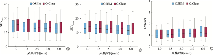
2. 客观评价:在L/B、SNR和SD方面(表 3),OSEM与Q. Clear均表现出相似的时间依赖性趋势。各时间组内Q. Clear的SD均低于OSEM,SNR高于OSEM。2 min Q. Clear的L/B、SNR及SD与6.0 min OSEM差异无统计学意义(P>0.05)。OSEM与Q. Clear在各时间组的SUVmax、SUVmean、肿瘤体积差异无统计学意义(P>0.05,图 2)。SUV重复性分析显示,两种重建算法在不同时间组的一致性极好,OSEM的ICC为0.943(SUVmax)和0.936(SUVmean),Q. Clear的ICC分别为0.951(SUVmax)和0.947(SUVmean);CV中位数为9.8%~11.6%,表明SUV在各时间组波动较小。
|
|
表 3 OSEM和Q. Clear图像客观参数(L/B、SNR和SD)对比[M(Q1, Q3)] Table 3 Comparison of objective parameters (L/B, SNR, and SD) between OSEM and Q. Clear images[M(Q1, Q3)] |
|
注:箱线图中箱体为25%~75% IQR范围,箱内实线为均值,须线延伸至1.5 IQR内极值。OSEM.有序子集最大期望值算法;Q. Clear. 贝叶斯惩罚似然算法;IQR.四分位距 图 2 OSEM和Q. Clear图像在不同采集时间的客观参数 A.最大标准摄取值(SUVmax);B.平均标准摄取值(SUVmean);C.病灶体积(LV) Figure 2 Objective parameters of OSEM and Q. Clear images A. Maximum standardized uptake(SUVmax); B. Mean standardized uptake(SUVmean); C. Lesion volume(LV) |
讨论
本研究系统评估了Q. Clear在68Ga-Pentixafor PET/MR肾上腺显像中的成像效果。结果表明,Q. Clear在2 min及以上采集时间能有效平衡噪声抑制与对比度保留,从而获取高质量图像。在保持6 min/床位采集时间不变的前提下,将68Ga-Pentixafor剂量减少至常规剂量的33.3%,Q. Clear能获得与OSEM全剂量相当的图像质量。
PET光子计数与放射性药物注射剂量成正比,决定了PET图像的质量,而患者所受辐射剂量与放射性药物注射剂量成正比。本研究结果基于Q. Clear在低计数条件下仍具备良好的噪声控制能力和对比度保留机制[17-20],能保证PA患者在大幅降低辐射剂量的同时,获得满足临床诊断需求的图像质量,与Kirchner等[21]的研究一致。在缩短检查时间、减少患者移动伪影方面具有潜在优势。
本研究结果显示,2 min Q. Clear图像的主客观评价与全剂量OSEM相当,满足临床需求,较既往weiwei等[12]的研究结果更优,差异可能是研究的β值不同。本研究的β值基于既往68Ga药物研究设置为1 000[11, 17-18],β值对图像质量具有双重效应:随着β值增高,噪声抑制能力随之增强,但可能降低部分对比度,导致病灶边缘的过度平滑,从而低估SUV值或遗漏微小病灶[22-23]。1、1.5 min OSEM和1 min Q. Clear的病灶显示程度显著低于6 min OSEM,提示过短的采集时间会削弱OSEM和Q. Clear的稳定性,图像噪声增大,干扰诊断。6 min Q. Clear图像整体质量主观评分较3 min Q. Clear降低,这可能与Q. Clear的惩罚函数特性相关:Q. Clear在短采集时间下优先保留高对比度信息,从而弥补计数不足导致的L/B和SNR下降[20]。
本研究仍存在一定局限性。首先,样本量相对较小,肥胖患者仅5例,占比较少(14%),结论外推至肥胖人群需谨慎,后续需扩大样本进行验证;其次,未对β值的优化进行系统性研究,未来可结合药物注射剂量、病灶体积和患者BMI动态调整β值,探索PA伴肥胖患者的个体化扫描方案[24-27];最后,对于68Ga-Pentixafor,缩短采集时间并不完全等效于降低药物剂量:前者主要引入统计噪声,后者伴随呼吸运动伪影增加及药代动力学改变。因此,Q. Clear在真实低剂量68Ga-Pentixafor场景下的效能仍有待前瞻性验证。
综上所述,在PA患者肾上腺趋化因子受体显像中,Q. Clear可在6 min/床位的采集条件下,将68Ga-Pentixafor剂量减少至33.3%,实现与全剂量OSEM相当的图像质量,为制定低剂量扫描方案提供了有力支持。
利益冲突 无
作者贡献声明 林华负责实验设计和论文撰写;李莉、胡云龙协助数据分析;苏鸣岗、李真林、夏春潮指导研究和论文修改;彭婉琳协助论文修改
| [1] |
Aline SM, Van LT, Sofie C, et al. Selective PET imaging of CXCR4 using the Al18F-labeled antagonist LY2510924[J]. Eur J Nucl Med Mol Imaging, 2025, 52(5): 1723-1738. DOI:10.1007/s00259-024-07025-w |
| [2] |
Zhang T, Chen T, Zhang W, et al. Clinical diagnosis and treatment of a patient with hypertension, hypokalemia, and bilateral adrenal adenomas[J]. Front Endocrinol (Lausanne), 2025, 16: 1558841. DOI:10.3389/fendo.2025.1558841 |
| [3] |
Zanotti-Fregonara P, Laforest R, Wallis JW. Fetal radiation dose from 18F-FDG in pregnant patients imaged with PET, PET/CT, and PET/MR[J]. J Nucl Med, 2015, 56(8): 1218-1222. DOI:10.2967/jnumed.115.157032 |
| [4] |
李进丹, 陈龙, 杨聪慧, 等. 低剂量PET/CT及PET/MRI临床研究进展[J]. 中国医学影像技术, 2021, 37(11): 1740-1743. Li JD, Chen L, Yang CH, et al. Clinical research progress of low-dose PET/CT and PET/MRI[J]. Chin J Med Imaging Technol, 2021, 37(11): 1740-1743. DOI:10.13929/j.issn.1003-3289.2021.11.035 |
| [5] |
Ribeiro D, Hallett W, Howes O, et al. Assessing the impact of different penalty factors of the Bayesian reconstruction algorithm Q. Clear on in vivo low count kinetic analysis of 11C PHNO brain PET-MR studies[J]. EJNMMI Res, 2022, 12(1): 11. DOI:10.1186/s13550-022-00883-1 |
| [6] |
Teoh EJ, McGowan DR, Schuster DM, et al. Bayesian penalised likelihood reconstruction (Q. Clear) of 18F-fluciclovine PET for imaging of recurrent prostate cancer: Semi-quantitative and clinical evaluation[J]. Br J Radiol, 2018, 91(1085): 20170727. DOI:10.1259/bjr.20170727 |
| [7] |
Roef MJ, Rijnsdorp S, Brouwer C, et al. Evaluation of quantitative Ga-68 PSMA PET/CT repeatability of recurrent prostate cancer lesions using both OSEM and bayesian penalized likelihood reconstruction algorithms[J]. Diagnostics (Basel), 2021, 11(6). DOI:10.3390/diagnostics11061100 |
| [8] |
Chicheportiche A, Goshen E, Godefroy J, et al. Can a penalized-likelihood estimation algorithm be used to reduce the injected dose or the acquisition time in 68Ga-DOTATATE PET/CT studies?[J]. EJNMMI Phys, 2021, 8(1): 13. DOI:10.1186/s40658-021-00359-6 |
| [9] |
Texte E, Gouel P, Thureau S, et al. Impact of the Bayesian penalized likelihood algorithm (Q. Clear ®) in comparison with the OSEM reconstruction on low contrast PET hypoxic images[J]. EJNMMI Phys, 2020, 7(1): 28. DOI:10.1186/s40658-020-00300-3 |
| [10] |
Rijnsdorp S, Roef MJ, Arends AJ. Impact of the noise penalty factor on quantification in Bayesian penalized likelihood (Q. Clear) reconstructions of 68Ga-PSMA PET/CT Scans[J]. Diagnostics (Basel), 2021, 11(5): 847. DOI:10.3390/diagnostics11050847 |
| [11] |
Carib PRRV, Koole M, D'Asseler Y, et al. Noise reduction using a Bayesian penalized-likelihood reconstruction algorithm on a time-of-flight PET-CT scanner[J]. EJNMMI Phys, 2019, 6(1): 22. DOI:10.1186/s40658-019-0264-9 |
| [12] |
Weiwei R, Chunxia Q, Fang L, et al. Q. Clear reconstruction for reducing the scanning time for 68Ga-DOTA-FAPI-04 PET/MR imaging[J]. Eur J Nucl Med Mol Imaging, 2023, 50(7): 1851-1860. DOI:10.1007/s00259-023-06134-2 |
| [13] |
陈芃灏, 符乙敏, 赵睿玥, 等. 探究64Cu-FAPI-XT PET/CT不同采集条件对图像质量的影响[J]. 肿瘤影像学, 2024, 33(4): 376-383. Chen QH, Fu YM, Zhao RY, et al. Investigation of the impact of different acquisition conditions on image quality in 64Cu-FAPI-XT PET/CT[J]. Tumor Imaging, 2024, 33(4): 376-383. DOI:10.19732/j.cnki.2096-6210.2024.04.006 |
| [14] |
Chaudhari AS, Mittra E, Davidzon GA, et al. Low-count whole-body PET with deep learning in amulticenter and externally validated study[J]. NPJ Digit Med, 2021, 4(1): 127. DOI:10.1038/s41746-021-00497-2 |
| [15] |
Driessen J, Zwezerijnen GJC, Schöder H, et al. The impact of semiautomatic segmentation methods on metabolic tumor volume, intensity, and dissemination radiomics in 18F-FDG PET scans of patients with classical hodgkin lymphoma[J]. J Nucl Med, 2022, 63(9): 1424-1430. DOI:10.2967/jnumed.121.263067 |
| [16] |
Eertink JJ, Pfaehler EAG, Wiegers SE, et al. Quantitative radiomics features in diffuse large B-cell lymphoma: Does segmentation method matter?[J]. J Nucl Med, 2022, 63(3): 389-395. DOI:10.2967/jnumed.121.262117 |
| [17] |
Franco MD, Fortunati E, Zanoni L, et al. β1600 Q. Clear digital reconstruction of [68Ga] Ga-DOTANOC PET/CT improves image quality in NET patients[J]. J Clin Med, 2024, 13(13): 3841. DOI:10.3390/jcm13133841 |
| [18] |
Xinchi H, Hillgan M, Esquinas Pedro L, et al. Impact of image reconstruction method on dose distributions derived from 90Y PET images: Phantom and liver radioembolization patient studies[J]. Phys Med Biol, 2020, 65(21): 215022. DOI:10.1088/1361-6560/aba8b5 |
| [19] |
Zanoni L, Argalia G, Fortunati E, et al. Can Q. Clear reconstruction be used to improve [68 Ga] Ga-DOTANOC PET/CT image quality in overweight NEN patients?[J]. Eur J Nucl Med Mol Imaging, 2022, 49(5): 1607-1612. DOI:10.1007/s00259-021-05592-w |
| [20] |
Sartoretti T, Skawran S, Gennari AG, et al. Fully automated computational measurement of noise in positron emission tomography[J]. Eur Radiol, 2024, 34(3): 1716-1723. DOI:10.1007/s00330-023-10056-w |
| [21] |
Kirchner J, O'Donoghue JA, Becker AS, et al. Improved image reconstruction of 89Zr-immunoPET studies using a Bayesian penalized likelihood reconstruction algorithm[J]. EJNMMI Phys, 2021, 8(1): 6. DOI:10.1186/s40658-021-00352-z |
| [22] |
Riet JT, Rijnsdorp S, Roef MJ, et al. Evaluation of a Bayesian penalized likelihood reconstruction algorithm for low-count clinical 18F-FDG PET/CT[J]. EJNMMI Phys, 2019, 6(1): 32. DOI:10.1186/s40658-019-0262-y |
| [23] |
Teoh EJ, McGowan DR, Macpherson RE, et al. Phantom and clinical evaluation of the Bayesian penalized likelihood reconstruction algorithm Q. Clear on an LYSO PET/CT system[J]. J Nucl Med, 2015, 56(9): 1447-1452. DOI:10.2967/jnumed.115.159301 |
| [24] |
Trägårdh E, Minarik D, Almquist H, et al. Impact of acquisition time and penalizing factor in a block-sequential regularized expectation maximization reconstruction algorithm on a Si-photomultiplier-based PET-CT system for 18F-FDG[J]. EJNMMI Res, 2019, 9(1): 64. DOI:10.1186/s13550-019-0535-4 |
| [25] |
Ribeiro D, Hallett W, Tavares AAS. Performance evaluation of the Q. Clear reconstruction framework versus conventional reconstruction algorithms for quantitative brain PET-MR studies[J]. EJNMMI Phys, 2021, 8(1): 41. DOI:10.1186/s40658-021-00386-3 |
| [26] |
Sadeghi F, Sheikhzadeh P, Farzanehfar S, et al. The effects of various penalty parameter values in Q. Clear algorithm for rectal cancer detection on 18F-FDG images using a BGO-based PET/CT scanner: a phantom and clinical study[J]. EJNMMI Phys, 2023, 10(1): 63. DOI:10.1186/s40658-023-00587-y |
| [27] |
Voert EEGWT, Muehlematter UJ, Delso G, et al. Quantitative performance and optimal regularization parameter in block sequential regularized expectation maximization reconstructions in clinical 68Ga-PSMA PET/MR[J]. EJNMMI Res, 2018, 8(1): 70. DOI:10.1186/s13550-018-0414-4 |