 2026, Vol. 46


随着我国居民生活水平的提高,人们对口腔健康及美观的需求日益提升,个体牙科医疗机构数量持续增长。牙科X射线机在临床应用越来越广泛[1-2]。牙科X射线机分口内机和口外机,口内机通常有固定式(如壁挂式、落地式等)、移动式以及便携式口内机,俗称便携式牙片机[3-4]。该类设备一般由电池供电,曝光开关直接位于设备上,临床使用时,操作人员通常站在受检者牙椅旁,单手或双手持握设备以保持其稳定并手动曝光,部分设备可搭配三脚架并利用遥控或电缆实现远程曝光。同室的操作人员会受到来自X射线管组件的泄漏辐射以及来自于受检者或检查室内其他物体的散射辐射[5-6],便携式牙片机特殊的使用模式使得同室操作人员和检查室内其他人员辐射安全问题成为研究热点[7-9]。本研究对某牙科诊所临床使用的便携式牙片机的辐射场剂量水平进行了检测,探讨便携式牙片机的实践正当性和辐射防护问题。
材料与方法1.检测设备:便携式牙科口内机按照外形分类可分为单手持握的“手枪式”和双手持握的“相机式”两类,按集光筒末端是否配置反散射屏蔽,可进一步分为带屏蔽型和无屏蔽型[7, 10]。本研究采用某国产便携式数字高频口腔X射线机,型号BLX-9,X射线管电压固定65 kV,标称管电流0.2 mA,曝光时间0.1~9.9 s连续可调,集光筒出口直径60 mm,主机重量1.8 kg。该设备属于“相机式”手持牙科机,未配置反散射屏蔽。
2.检测仪器:AT1123 X-γ射线巡测仪(白俄罗斯ATOMTEX公司),能量范围15 keV~10 MeV,短时曝光响应时间30 ms,短时曝光剂量率响应范围50 nSv/h~10 Sv/h。该仪器由中国计量科学研究院校准并检定合格,使用时在校准有效期内。
3.检测方法:将标准水模(30 cm × 30 cm × 20 cm)放在牙椅上模拟受检者体部,将标准CT剂量头模(直径16 cm,高15 cm)放置在水模上模拟受检者头部,头模中心距离地面105 cm。
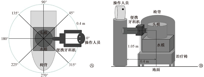
操作人员穿戴铅橡胶颈套和铅橡胶防护服,铅当量均为0.5 mm Pb(120 kV),未佩戴铅眼镜。模拟临床使用便携式牙片机的日常操作状态,位于受检者的右手边,双手持握设备、手臂自然弯曲,将便携式牙片机集光筒出口抵在头模表面,采用设备预设固定管电压和管电流,曝光时间设置为0.5 s,满足测量设备时间响应要求[11],主射束对准头模中心曝光。测量示意图示于图 1。
|
图 1 测量示意图 A.俯视图;B. 侧视图 Figure 1 Schematic diagram of the measurement setup A. Top view; B. Side view |
(1) 操作人员剂量水平:操作人员体表距离头模中心40 cm,参考WS 76-2020《医用X射线诊断设备质量控制检测规范》[12],按照透视防护区术者位测量关注点的高度要求,分别在便携式牙片机曝光时测量距地面分别为155、135、125、105、80和20 cm(分别指操作人员眼晶状体、甲状腺、胸部、腹部、性腺以及足部)的周围剂量当量率,同时测量操作人员持握便携式牙片机时左右手的手指、手背位置的周围剂量当量率,每个测量点重复测量两次,取其平均值,扣除本底后结合校准因子计算剂量结果。
(2) 个人防护用品屏蔽效果:分别测量操作人员铅橡胶颈套和防护服内,对应的甲状腺、胸部、腹部位置的周围剂量当量率水平。
(3) 辐射场剂量水平:以操作人员背后方向为0°,分别测量0°、45°、90°、135°、180°、225°、270°和315° 8个方向,距离头模中心1.0、1.5和2.0 m处,距地面高度为105 cm的周围剂量当量率,辐射场测量点位示于图 2。为进一步明确便携式牙片机操作场所周围剂量当量率水平以及对周围公众的潜在影响,本研究还测量了90°方向(垂直于主射线方向)上距离受检者4.0、6.0、10.0 m处的周围剂量当量率。

|
图 2 测量点位示意图 Figure 2 Schematic diagram of the radiation field measurement points |
结果
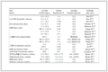
1.操作人员位置剂量水平:操作人员不同高度(部位)以及手部所在位置的周围剂量当量率列于表 1。由表 1可知,操作人员铅衣外剂量率范围为196.4~1 334.9 μSv/h,最高点位于距地面135 cm处(甲状腺部位),最低点位于距地面20 cm处(足部位置);手部剂量率范围为197.9~459.9 μSv/h。在0.5 mm Pb防护用品(铅橡胶颈套和防护服)内,不同位置的周围剂量当量率衰减比例均超过98%。
|
表 1 操作人员体部和手部位置的平均周围剂量当量率 Table 1 Mean ambient dose equivalent rate at the body and hand positions of the operator |
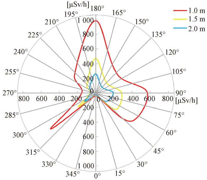
2.工作场所周围剂量当量率:以受检模体为中心,受检者右侧方向,即操作人员后背方向为0°,测量每45°方向上不同距离工作场所周围剂量当量率(图 3),具体结果列于表 2。操作人员背后(0°方向)和椅背方向(270°方向)剂量率较低,主射线束方向剂量较其他方向高。考虑到临床诊室测量条件,本研究测量了90°方向(垂直于主射线方向)的周围剂量当量率,发现在现场未设置屏蔽条件下距离受检者4、6和10 m处周围剂量当量率扣除本底后,分别为50.0、16.6、5.3 μSv/h。
|
图 3 等距离(1.0、1.5、2.0 m)的剂量曲线图 Figure 3 Doses curve at equal distances (1.0, 1.5, and 2.0 m) |
|
|
表 2 距离头模1.0、1.5和2.0 m处的不同方向的周围剂量当量率(μSv/h) Table 2 Ambient dose equivalent rates at distances of 1.0, 1.5, and 2.0 m in different directions within the radiation field centered on the head phantom (μSv/h) |
讨论
第一台便携式牙片机可以追溯到20世纪90年代初,最初被用于军事领域[8],之后广泛应用于灾害救援、犯罪现场以及兽医等[13-14],又逐渐延伸至在家中或在医疗机构中活动受限的牙科受检者。相对于隔室操作的人员,操作便携式牙片机的操作人员剂量较高,而且这类设备容易逃避监管。2015年,欧洲牙颌面放射学会(EADMFR)建议,便携式牙科X射线设备不得用于牙科诊所的常规牙科X射线检查,仅应在无法或不可能将受检者转移到固定安装的X射线设备的特定情况下使用[15-16]。欧洲辐射防护主管机构负责人协会(HERCA)的文件指出,此类设备仅应在传统固定式牙科设备无法运行的情况下使用,例如疗养院、居家养老机构或行动不便者之家,或用于法医牙科、军事行动等特殊场景[17]。我国也规定,包括便携式牙片机在内的便携式设备不应用于常规检查[18]。
国外已有研究者针对不同机型的便携式牙科口内机的辐射剂量特征开展了测试分析[19-21],而国内对于便携式牙片机操作人员受照剂量研究[22]和场所剂量水平研究相对匮乏。在Leadbeatter和Diffey[6]的研究中,无散射屏蔽的情况下,按每年拍片3 000次,操作人员的右手和眼部的年剂量分别为2.4和1.4 mSv;Goren等[19]报道,无散射屏蔽的情况下,按每年拍片15 000次,操作人员的胸部、眼和手的年剂量分别为0.18、0.225和0.45 mSv;Hosseini等[20]报道,无散射屏蔽的情况下,每次拍(上牙)片操作人员的全身、手指、眼晶状体和甲状腺的剂量分别为95、1 410、111和53 μSv;在Cho和Han[21]的研究中,无散射屏蔽的情况下,每次拍片操作人员的胸部、腰部和手的剂量分别为0.043、0.062和0.129 μSv。通过对比可以发现,不同研究中操作人员累积剂量并不一致,这是由于便携式牙片机的参数、防护配置、操作人员站立位置以及工作负荷都可能对辐射水平产生较大的影响[9]。Danforth等[23]测量了操作人员在使用某便携式牙科口内机时接受的剂量,结果显示年有效剂量为0.047 mSv。国内牙科放射学操作绝大部分为隔室操作,邓君等[24]报道,牙科放射学设备操作人员年有效剂量为0.326 mSv,远低于《电离辐射防护与辐射源安全基本标准》中的要求[25],但普遍高于国外研究报道,因此更应该关注同室操作便携式牙科机职业人员的辐射风险。同室操作设备工作场所剂量水平方面,宗睿等[26]和何丽娟等[27]分别测量了移动式头部锥形束CT(CBCT)和数字减影血管造影(DSA)工作场所辐射剂量分布情况,显示出业界对于同室操作人员辐射防护的关注。
在本研究中,操作人员使用全身和甲状腺防护用品对辐射的防护效果均在98%以上。Hu等[28]报道铅眼镜对眼晶状体剂量的屏蔽在75%以上,提示在同室操作的牙科放射学实践中,穿戴放射防护用品是十分必要的。国外研究报道,于X射线管集光筒末端配置反散射屏蔽,手部散射剂量屏蔽率可达70%,眼晶状体剂量屏蔽率为68%,操作人员年剂量降幅近90% [6, 21]。本研究所用的国产便携式牙片机未配置反散射屏蔽,我国相关标准中也未有类似规定。美国食品药品监督管理局规定,便携式X射线设备必须配备某种形式的反散射屏蔽[29],反散射屏蔽的最小规格应为铅当量0.25 mm Pb,直径为15.2 cm,并且能够定位在距离末端不超过1 cm的位置。
在诊断X射线能谱范围中,康普顿效应占主导地位,散射角对称地分布在光子入射角的两边[13]。本研究测量过程中,便携式口内机正对头模中心,X射线入射方向是由y轴负方向到y轴正方向。在没有其他物体的干扰情况下,散射辐射呈现关于y轴对称的分布特征。225°方向和135°方向射线受物体的干扰较小,近似y轴对称,也说明了散射辐射是周围辐射剂量的主要贡献源。但由于0°方向的操作人员以及270°方向的牙椅椅背存在,所以在这两个方向有明显变小,即射线被人体和椅背衰减,剂量率降低。180°方向的测量位置在操作人员的对面,此处的辐射是主射束穿透头模之后的辐射、部分散射辐射及泄漏辐射之和,周围剂量当量率最大。建议在操作便携式牙片机放射实践中,若助手无法离开现场,应避免处于正对X射线方向,应站立在椅背后或操作人员背后等剂量较低位置;根据本研究的测量结果,与主射束方向呈90°方向时,在未设置现场屏蔽的情况下,即便距离较远处的剂量率水平也高于固定式牙片机房屏蔽体外周围剂量率限值,因此在操作便携式牙片机时,应禁止公众和受检者家属靠近[30]。需要指出的是,本研究检测人员直接在受检者周边测量,检测人员本身也作为散射体会产生散射辐射,可能会使测得的结果偏高。
大部分便携式牙片机超过2 kg,某款便携式牙片机的重量甚至接近4 kg[31]。便携式牙片机普遍管电流较低,检查过程中为了获得诊断所需的曝光量,曝光时间需要长达数秒,在此过程中操作人员使用手持的方式操作难免会产生位移,进而产生伪影影响成像质量,因此便携式牙片机的图像质量可能会低于固定式设备[16]。
我国职业卫生标准《放射诊断放射防护要求》(GBZ 130-2020)[18]规定,移动式和便携式X射线设备不应用于常规检查。只有在不能实现或在医学上不允许把受检者送到固定设备进行检查的情况下,并在采取严格的防护措施后,才能使用移动式或便携式X射线设备在床旁操作,实施医学影像检查。我国对医疗机构使用便携式牙片机监管较严,但部分医疗机构仍然存在不合规使用便携式牙片机的情况。
本研究测量了便携式牙片机使用场所不同方向不同距离的剂量水平分布情况,为同室操作人员站位提供参考,并提出部分放射防护建议。目前,我国尚无便携式牙片机实践正当性与防护最优化的专项指南,建议可参考国际上已有的牙科X射线设备放射防护指南[32-33],再基于国内便携式牙片机的实际情况,尽快建立针对性较强的防护标准或防护共识,明确规范便携式牙片机的使用范围、技术参数、反散射屏蔽配置以及防护要求等,保护职业人员健康权益。
利益冲突 无
作者贡献声明 曹金平、刘国庆负责现场测量、数据分析和论文撰写;徐辉负责选题设计和指导;岳保荣、赵俏俏参与论文修改
| [1] |
Public Health England (PHE) and Faculty of General Dental Practice (UK) (FGDP[UK]). Guidance notes for dental practitioners on the safe use of X-ray equipment. 2nd ed[M]. London: PHE and FGDP(UK), 2020.
|
| [2] |
Lommen J, Schorn L, Nitschke J, et al. Accuracy in positioning of dental X-ray images - a comparative study of a portable X-ray device and a wall-mounted device[J]. Saudi Dent J, 2021, 33(8): 884-889. DOI:10.1016/j.sdentj.2021.09.018 |
| [3] |
Ohtani M, Oshima T, Mimasaka S. Extra-oral dental radiography for disaster victims using a flat panel X-ray detector and a hand-held X-ray generator[J]. J Forensic Odontostomatol, 2017, 2(35): 28-34. |
| [4] |
Gray JE, Bailey ED, Ludlow JB. Dental staff doses with handheld dental intraoral x-ray units[J]. Health Phys, 2012, 102(2): 137-142. DOI:10.1097/HP.0b013e318230778a |
| [5] |
Smith R, Tremblay R, Wardlaw GM. Evaluation of stray radiation to the operator for five hand-held dental X-ray devices[J]. Dentomaxillofac Radiol, 2019, 48(5): 20180301. DOI:10.1259/dmfr.20180301 |
| [6] |
Leadbeatter J, Diffey J. Evaluation of radiation exposure to operators of portable hand-held dental X-ray units[J]. Phys Eng Sci Med, 2021, 44(2): 377-385. DOI:10.1007/s13246-021-00985-5 |
| [7] |
Ramesh DV, Wale M, Thriveni R, et al. Hand-held X-ray device: a review[J]. Indian Acad Oral Med Radiol, 2018, 30: 153-157. DOI:10.4103/jiaomr.jiaomr_36_18 |
| [8] |
Van Dis ML, Miles DA, Parks ET, et al. Information yield from a hand-held dental x-ray unit[J]. Oral Surg Oral Med Oral Pathol, 1993, 76(3): 381-385. DOI:10.1016/0030-4220(93)90272-6 |
| [9] |
Makdissi J, Pawar RR, Johnson B, et al. The effects of device position on the operator's radiation dose when using a handheld portable X-ray device[J]. Dentomaxillofac Radiol, 2017, 45(3): 20150245. DOI:10.1259/dmfr.20150245 |
| [10] |
Public Health England (PHE). Phecrce-023. Guidance on the safe use of hand-held dental X-ray equipment[R]. London: PHE, 2016.
|
| [11] |
徐辉, 赵锡鹏, 岳保荣, 等. RJ32-2106P和AT1123两种Χ-γ剂量率仪响应时间性能研究[J]. 中国辐射卫生, 2018, 27(6): 595-598. Xu H, Zhao XP, Yue BR, et al. Study on response time performance of RJ32-2106P and AT1123 X-γ ray dose rate meters[J]. Chin J Radiol Health, 2018, 27(6): 595-598. DOI:10.13491/j.issn.1004-714X.2018.06.021 |
| [12] |
国家卫生健康委员会. WS 76-2020医用X射线诊断设备质量控制检测规范[S]. 北京: 中国标准出版社, 2020. National Health Commission. WS 76-2020 Specification for testing of quality control in medical X-ray diagnostic equipment[S]. Beijing: Standards Press of China, 2020. |
| [13] |
Rottke D, Gohlke L, Schrödel R, et al. Operator safety during the acquisition of intraoral images with a handheld and portable X-ray device[J]. Dentomaxillofac Radiol, 2018, 47(3): 20160410. DOI:10.1259/dmfr.20160410 |
| [14] |
Hermsen KP, Jaeger SS, Jaeger MA. Radiation safety for the NOMAD portable X-ray system in a temporary morgue setting[J]. J Forensic Sci, 2008, 53(4): 917-921. DOI:10.1111/j.1556-4029.2008.00766.x |
| [15] |
Geist JR. Handheld intraoral dental x-ray devices should supplement but not replace conventional radiographic equipment[J]. Oral Surg Oral Med Oral Pathol Oral Radiol, 2021, 132(3): 257-259. DOI:10.1016/j.oooo.2021.06.001 |
| [16] |
Berkhout WE, Suomalainen A, Brüllmann D, et al. Justification and good practice in using handheld portable dental X-ray equipment: a position paper prepared by the European Academy of Dentomaxillofacial Radiology (EADMFR)[J]. Dentomaxillofac Radiol, 2015, 44(6): 20140343. DOI:10.1259/dmfr.20140343 |
| [17] |
Heads of the European Radiological Protection Competent Authorities. Position statement on use of handheld portable dental x-ray equipment[R]. Vilnius: HERCA, 2014.
|
| [18] |
国家卫生健康委员会. GBZ 130-2020放射诊断放射防护要求[S]. 北京: 中国标准出版社, 2020. National Health Commission. GBZ 130-2020 Requirements for radiological protection in diagnostic radiology[S]. Beijing: Standards Press of China, 2020. |
| [19] |
Goren AD, Bonvento M, Biernacki J, et al. Radiation exposure with the NOMAD portable X-ray system[J]. Dentomaxillofac Radiol, 2008, 37(2): 109-112. DOI:10.1259/dmfr/33303181 |
| [20] |
Hosseini Pooya SM, Hafezi L, Manafi F, et al. Assessment of the radiological safety of a Genoray portable dental X-ray unit[J]. Dentomaxillofac Radiol, 2015, 44(3): 20140255. DOI:10.1259/dmfr.20140255 |
| [21] |
Cho JY, Han WJ. The reduction methods of operator's radiation dose for portable dental X-ray machines[J]. Restor Dent Endod, 2012, 37(3): 160-164. DOI:10.5395/rde.2012.37.3.160 |
| [22] |
黄丽华, 林春培, 黄海潮, 等. 国产MSD型牙科X射线机的防护改造[J]. 中华放射医学与防护杂志, 2001, 21(3): 212-214. Huang LH, Lin CP, Huang HC, et al. Protection modification of domestic MSD dental X-ray machine[J]. Chin J Radiol Med Prot, 2001, 21(3): 212-214. DOI:10.3760/cma.j.issn.0254-5098.2001.03.032 |
| [23] |
Danforth RA, Herschaft EE, Leonowich JA. Operator exposure to scatter radiation from a portable hand-held dental radiation emitting device (Aribex NOMAD) while making 915 intraoral dental radiographs[J]. J Forensic Sci, 2009, 54(2): 415-421. DOI:10.1111/j.1556-4029.2008.00960.x |
| [24] |
邓君, 王拓, 范胜男, 等. 2015年我国医用放射工作人员职业外照射个人剂量水平与分析[J]. 中国辐射卫生, 2017, 26(4): 398-400. Deng J, Wang T, Fan SN, et al. Results and analysis on individual dose level of occupational external exposure to radiation for medical radiation workers in China in 2015[J]. Chin J Radiol Health, 2017, 26(4): 398-400. DOI:10.3969/j.issn.1004-714X.2017.04.004 |
| [25] |
国家质量监督检验检疫总局. GB 18871-2002电离辐射防护与辐射源安全基本标准[S]. 北京: 中国标准出版社, 2003. General Administration of Quality Supervision, Inspection and Quarantine of the People's Republic of China. GB 18871-2002 Basic standards for protection against ionizing radiation and for the safety of radiation sources[S]. Beijing: Standards Press of China, 2003. |
| [26] |
宗睿, 王楠, 薄文柱, 等. 移动式头部锥形束CT(CBCT)周围剂量场分布测试[J]. 中国辐射卫生, 2021, 30(5): 638-642. Zong R, Wang N, Bo WZ, et al. Measurement of dose field distribution around mobile cone beam computed tomography (CBCT)[J]. Chin J Radiol Health, 2021, 30(5): 638-642. DOI:10.13491/j.issn.1004-714X.2021.05.024 |
| [27] |
何丽娟, 张岭, 何廷贵, 等. 基于PBU-60体模的介入患者及职业人员辐射剂量学研究[J]. 中国辐射卫生, 2021, 30(4): 417-422, 427. He LJ, Zhang L, He TG, et al. Radiation dosimetric study of patients and professionals in an intervention procedure based on PBU-60 anthropomorphic phantom[J]. Chin J Radiol Health, 2021, 30(4): 417-422, 427. DOI:10.13491/j.issn.1004-714X.2021.04.006 |
| [28] |
Hu P, Kong Y, Chen B, et al. Shielding effect of lead glasses on radiologists' eye lens exposure in interventional procedures[J]. Radiat Prot Dosim, 2017, 174(1): 136-140. DOI:10.1093/rpd/ncw098 |
| [29] |
Center for Devices and Radiological Health. Guidance for industry and FDA staff, radiation safety considerations for X-ray equipment designed for hand-held use[R]. Maryland: CDRH, 2008.
|
| [30] |
Health Canada. Radiation protection in dentistry - recommended safety procedures for the use of dental X-ray equipment - safety code 30[S]. Ottawa: Health Canada, 2022.
|
| [31] |
Pittayapat P, Oliveira-Santos C, Thevissen P, et al. Image quality assessment and medical physics evaluation of different portable dental X-ray units[J]. Forensic Sci Int, 2010, 201(1-3): 112-117. DOI:10.1016/j.forsciint.2010.04.041 |
| [32] |
Australian Radiation Protection and Nuclear Safety Agency. Radiation protection series No 10. Code of practice and safety guide for radiation protection in dentistry[R]. Victoria: Chief Executive Officer of ARPANSA, 2005.
|
| [33] |
Lurie AG, Kantor ML. Contemporary radiation protection in dentistry: recommendations of National Council on Radiation Protection and Measurements Report No. 177[J]. J Am Dent Assoc, 2020, 151(10): 716-719.e3. DOI:10.1016/j.adaj.2020.05.007 |