 2025, Vol. 45


2. 山西白求恩医院(山西医学科学院 同济山西医院)山西医科大学第三医院肿瘤中心,太原 030032
2. Department of Oncology, Shanxi Bethune Hospital, Shanxi Academy of Medical Sciences, Tongji Shanxi Hospital, Third Hospital of Shanxi Medical University, Taiyuan 030032, China
宫颈癌是我国高发的妇科恶性肿瘤,其治疗策略依据疾病分期而定。局部晚期患者以放射治疗为主要根治手段,存在高危因素的早中期患者则需将放射治疗作为重要的术后辅助手段[1-2]。螺旋断层放射治疗系统(helical tomotherapy,HT,简称Tomo系统)是当前先进的放射治疗设备之一,通过机架360°旋转照射及气动多叶准直器(MLC)在每个投影内打开时间的调节实现束流强度调制,能对复杂形状的靶区进行精准调强治疗[3]。在宫颈癌放射治疗中,相较于传统直线加速器,Tomo系统在提升靶区剂量均匀性和一致性、降低重要危及器官剂量方面更具优势[4-5],但因其子野数目多、计划复杂度高,对剂量验证的要求也更为严格[6]。
ArcCheck是常见的三维剂量验证设备,具有实际角度测量、操作简便、结果稳定等优点,但其21 cm的有效测量长度存在局限[7-14]。对于靶区包含腹膜后淋巴结的宫颈癌患者,计划靶区(PTV)长度可能超过21 cm,若直接采用常规等中心摆位验证可能照射到ArcCheck的电子元件。目前解决这一问题的常用方法有两种:一是移动摆位中心单次测量,可能因范围不全影响γ通过率[15];二是利用ArcCheck的合并(merge)功能分两段测量后拼接,但存在操作复杂、工作量大的问题[16-17]。
本研究通过分析移动摆位中心和分段拼接两种测量方式下宫颈癌超长靶区验证通过率的分布情况,旨在为临床验证工作提供参考依据。
资料与方法1.病例选择:本研究回顾性纳入2021年2月至2024年5月于山西白求恩医院肿瘤放射治疗中心接受Tomo系统放射治疗的24例宫颈癌超长靶区患者。这些患者靶区长度为23.5 ~ 36.0 cm,平均29.5 cm。治疗计划射野宽度(FW)设为2.5 cm,螺距(pitch)值为0.287,调制因子(MF)在2.0 ~ 2.8范围内。
2.设备:本研究治疗设备为螺旋断层治疗系统Tomotherapy H(美国安科锐公司),计划制定采用Tomo计划系统5.1.8.23版本。剂量验证设备为ArcCheck(美国Sun Nuclear公司),其为圆柱形三维剂量验证设备,圆柱表面2.9 cm深度处有1 386个半导体探头。验证时使用PMMA CavityPlug插件(美国Sun Nuclear公司),配套分析软件为SNC Patient 8.4.1版本。
3.移动摆位中心单独测量:在治疗质量保证(DQA)工作站,将患者治疗计划移植至实际扫描的ArcCheck模体。移动模体,使其在不照射电子元件前提下,测量范围尽可能大且靶区位于模体中心。计算剂量分布并以DICOM格式导出至SNC Patient软件。测量时,调取验证计划,红激光对准模体摆位中心。计算测量结果与验证计划计算结果的γ通过率。
4.分段拼接的测量
(1) 50 cm虚拟模体的导入:因ArcCheck有效测量范围仅21 cm,无法单次完整计算超长靶区,需导入厂家50 cm虚拟模体。利用DICOM导入程序(Dicom Push),将Patient Name命名为_phantom,系统自动识别图像为模体图像;指定ArcCheck质量密度(IVDT)曲线,其中Hu值-1 024、-1 000和215的对应密度值为0、0.001和1.183,分别对应零点、空气及模体均匀介质的Hu值和密度值。
(2) 验证计划的计算和导出:在DQA工作站,将患者治疗计划移植至50 cm虚拟模体,使靶区位于模体中心,计算剂量分布并以DICOM格式导出至SNC Patient软件。
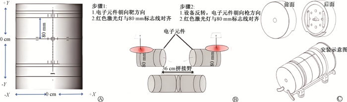
(3) 验证计划的测量:①将ArcCheck模体电子元件朝向机架孔径外部,调取验证计划,按模体摆位中心摆位后,移动治疗床使红色激光灯与80 mm标志线对齐;出束测量,在SNC Patient软件“CAX Offset”选项Y方向输入80 mm,保存测量文件命名为“80 mm”。②将模体反转,电子元件朝向机架孔径内部,调取验证计划,按模体摆位中心摆位后,移动治疗床使红色激光灯重新与80 mm标志线对齐;重新出束测量,在SNC “CAX Offset”选项Y方向输入-80 mm,勾选“ArcCheck Inverted”选项,保存测量文件命名为“-80 mm”。③在菜单栏选“工具>组合> SNC设备数据”,选择上述两测量文件,融合生成拼接文件,命名为“merge”,计算其与使用50 cm虚拟模体的验证计划计算结果的γ通过率。ArcCheck摆位中心、80 mm标志线见图 1A,具体摆位见图 1B。
|
图 1 ArcCheck模体及摆位、散射块安装示意图[18-19] A. ArcCheck模体摆位中心、80 mm标记线;B. ArcCheck拼接测量摆位;C. 散射块安装示意图 Figure 1 Schematic diagrams of ArcCheck phantom and its setup and scattering block installation [18-19] A. Isocenter and 80 mm reference line of the ArcCheck phantom; B. Setup of the ArcCheck phantom for segmented measurements combined with merging; C. Installation of scattering blocks |
(4) 安装散射块后验证计划的测量:将ArcCheck模体安装散射块,具体安装见示意图 1C。按步骤(3)重新测量,模体正反情况下测量文件分别命名为“80 mmS”和“-80 mmS”;生成拼接文件命名为“mergeS”,计算其与使用50 cm虚拟模体的验证计划计算结果的γ通过率。
(5) 安装散射块前后分段测量文件γ通过率的计算:分别计算ArcCheck模体安装散射块前后的分段测量文件“80 mm”“80 mmS”与使用50 cm虚拟模体的验证计划计算结果的γ通过率。计算时,先打开测量文件,再打开计算文件,SNC Patient软件会据测量文件探测范围自动调整计算文件范围。
5.评价标准:本研究以阈值水平10%、绝对剂量(全局)、相对剂量的3%/2 mm、2%/2 mm和1%/1 mm的γ通过率作为评价标准。
6.统计学处理:采用SPSS 19.0软件进行统计学分析。利用K-S检验数据正态性,以中位数和四分位数间距[M(Q1,Q3)]表示γ通过率。对移动摆位中心单独测量与不加散射块的分段测量后拼接、移动摆位中心单独测量与加散射块的分段测量后拼接两种方式的γ通过率分别进行配对Wilcoxon检验;同时,对ArcCheck模体安装散射块前后的分段测量文件“80 mm”“80 mmS”的γ通过率进行配对Wilcoxon检验。P < 0.05为差异具有统计学意义。
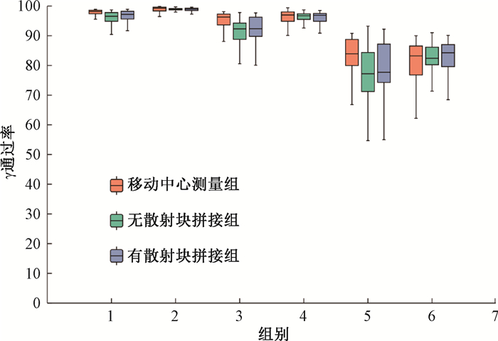
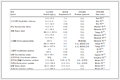
结果1.移动摆位中心单独测量与加散射块、不加散射块拼接文件γ通过率的比较:如图 2所示,在不同γ通过率标准下,移动摆位中心单独测量的验证结果均高于加或不加散射块的拼接方式。然而,经统计学分析,移动摆位中心单独测量与加散射块拼接测量在各评价标准下的γ通过率差异均无统计学意义(P>0.05);与不加散射块拼接方式相比,在3%/2 mm绝对剂量(全局)标准下,γ通过率差异具有统计学意义(Z=-2.31,P=0.02),其余评价标准的统计学结果列于表 1。
|
注:1、3、5分别为3%/2 mm、2%/2 mm、1%/1 mm绝对剂量;2、4、6分别为3%/2 mm、2%/2 mm、1%/1 mm相对剂量 图 2 移动摆位中心单独测量组与加散射块、不加散射块拼接组γ通过率的分布情况 Figure 2 Distributions of γ-pass rates under individual measurements by moving the phantom isocenter and segmented measurements with and without scattering blocks |
|
表 1 宫颈癌超长靶区不同方式测量时γ通过率的比较 Table 1 Comparison of γ-pass rates for different measurement methods of ultra-long target volumes in cervical cancer |
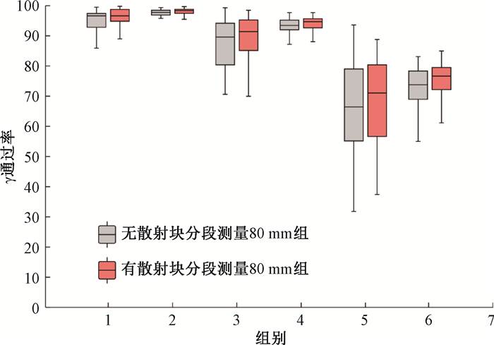
2.ArcCheck模体安装散射块前后的分段测量文件γ通过率的比较:如图 3所示,加散射块可提高分段测量的验证通过率。在3%/2 mm相对剂量标准下,γ通过率差异具有统计学意义(Z=-2.11,P=0.04),其余评价标准的统计学结果也列于表 1。
|
注:AD. 绝对剂量;RD. 相对剂量注:1、3、5分别为3%/2 mm、2%/2 mm、1%/1 mm绝对剂量;2、4、6分别为3%/2 mm、2%/2 mm、1%/1 mm相对剂量 图 3 加散射块、不加散射块分段测量文件γ通过率的分布情况 Figure 3 Distribution of γ-pass rates for segmented measurement files with and without scattering blocks |
讨论
ArcCheck是一种广泛使用的三维剂量验证设备,但其有效测量长度仅为21 cm。当靶区长度超出此范围时,常规等中心摆位的验证方法可能会照射到设备的电子元件部分,导致设备损伤,进而影响剂量的准确性。周本梅等[15] 的研究发现,当靶区长度大于ArcCheck的有效测量长度时,可将ArcCheck沿出床方向平移5 cm,以降低照射电子元件的风险。同时,在3%/3 mm和3%/2 mm的评价标准下,超长靶区组的γ通过率仍表现良好。此外,宫颈癌常规靶区组Short-0 cm(测量全部靶区)和Short-5 cm(摆位中心移动5 cm只测量部分靶区)两组的γ通过率差异具有统计学意义,这表明模体摆位中心的偏移与靶区长度均会对剂量通过率产生影响。对于长度超过21 cm的靶区,虽然无法使用ArcCheck单次测量完整靶区长度,但厂家提供的分段测量后拼接的方式可将检测范围延长至36 cm,从而模拟超长靶区的完整测量。本研究对比了宫颈癌超长靶区移动摆位中心单独测量(在不照射元器件的前提下,使测量范围尽可能大)与分段测量拼接方式的γ通过率,结果显示,移动摆位中心单独测量与加散射块的拼接测量在各种评价标准下的γ通过率差异均无统计学意义。相较于拼接测量对软件版本和物理师要求较高、操作流程复杂、剂量验证工作量大等一系列问题,移动摆位中心单独测量的方法更适合临床工作需求。
ArcCheck模体可在一端安装长为9.1 cm的高密度聚乙烯散射块,用于在测量超出模体边缘的靶区时考虑散射和二次辐射的贡献。安装散射块后,21 cm × 40 cm 6 MV射野的散射辐射损失导致的测量误差为2% 或更小(相对于完全考虑散射辐射的几何结构)[19]。本研究比较了ArcCheck安装散射块前后的分段测量的验证通过率结果,发现加散射块可提高γ通过率,且差异具有统计学意义。原因在于验证计划的计算结果是基于50 cm虚拟模体,即相对于完全考虑散射辐射的几何结构;而分段测量时安装散射块减少了因散射辐射损失导致的测量误差。然而,对于采用拼接方式后的γ通过率,是否加散射块的影响较小,本研究未发现两者γ通过率的差异具有统计学意义。这可能是由于剂量拼接过程中对重叠区域取平均值,从而减少了因散射辐射损失导致的测量误差。尽管如此,本研究在比较移动摆位中心单独测量与加或不加散射块的拼接测量时发现,移动摆位中心单独测量的γ通过率高于不加散射块的拼接测量,在3%/2 mm绝对剂量(全局)标准下,γ通过率的差异具有统计学意义。基于以上发现,如果在临床中使用分段测量后拼接的方式对超长靶区进行剂量验证,建议安装散射块进行测量。
在Tomo设计验证计划时,可以选择将ArcCheck模体移动,使靶区位于模体中心生成中心型验证计划,也可以使靶区位于模体半导体探测器上生成偏心型验证计划。本研究所有剂量验证均采用中心型验证计划。原因在于岳麒等 [20] 的研究指出,ArcCheck应用于Tomo验证的中心型验证方式比偏心型验证方式对叶片打开时间误差的灵敏度更高,对误差的检测能力更强,且中心型验证计划在不同标准下的γ分析结果的相关性均强于偏心型验证计划。
使用移动摆位中心单独测量的方式验证超长靶区时,能否通过增加散射块提高验证通过率,将在今后的研究中继续探讨。此外,本研究的剂量验证测量均在Tomo系统进行,今后还需在常规直线加速器上进一步验证。
综上所述,使用ArcCheck测量宫颈癌超长靶区时,建议优先采用移动摆位中心单独测量的方式。若需使用分段测量进行拼接,则应在测量时安装散射块,以提高验证通过率,确保剂量验证的准确性。
利益冲突 无
作者贡献声明 雷俊杰负责实验的实施及论文撰写;刘济人负责实验数据的统计学分析;潘孝鹏、侯旭负责剂量验证数据的采集;金晓丽负责实验设计、组织实施及论文修改
| [1] |
Sung H, Ferlay J, Siegel RL, et al. Global cancer statistics 2020: GLOBOCAN estimates of incidence and mortality worldwide for 36 cancers in 185 countries[J]. CA Cancer J Clin, 2021, 71(3): 209-249. DOI:10.3322/caac.21660 |
| [2] |
Huang K, Jia M, Li P, et al. Radiotherapy improves the survival of patients with metastatic cervical cancer: a propensity-matched analysis of SEER database[J]. Int J Gynecol Cancer, 2018, 28(7): 1360-1368. DOI:10.1097/IGC.0000000000001313 |
| [3] |
Mackie TR, Holmes T, Swerdloff S, et al. Tomotherapy: a new concept for the delivery of dynamic conformal radiotherapy[J]. Med Phys, 1993, 20(6): 1709-1719. DOI:10.1118/1.596958 |
| [4] |
Li D, Wang D, Feng S, et al. Comparing dosimetric and cancer control outcomes after intensity-modulated radiation therapy and tomotherapy for advanced cervical cancer[J]. Oncol Lett, 2022, 24(1): 239. DOI:10.3892/ol.2022.13359 |
| [5] |
Cheng YK, Kuo SH, Yen HH, et al. The prognostic significance of pretreatment squamous cell carcinoma antigen levels in cervical cancer patients treated by concurrent chemoradiation therapy and a comparison of dosimetric outcomes and clinical toxicities between tomotherapy and volumetric modulated arc therapy[J]. Radiat Oncol, 2022, 17(1): 91. DOI:10.1186/s13014-022-02063-w |
| [6] |
Cavinato S, Fusella M, Paiusco M, et al. Quantitative assessment of helical tomotherapy plans complexity[J]. J Appl Clin Med Phys, 2023, 24(1): e13781. DOI:10.1002/acm2.13781 |
| [7] |
Thiyagarajan R, Nambiraj A, Sinha SN, et al. Analyzing the performance of ArcCHECK diode array detector for VMAT plan[J]. Rep Pract Oncol Radiother, 2016, 21(1): 50-56. DOI:10.1016/j.rpor.2015.10.004 |
| [8] |
Steers JM, Fraass BA. IMRT QA and gamma comparisons: the impact of detector geometry, spatial sampling, and delivery technique on gamma comparison sensitivity[J]. Med Phys, 2021, 48(9): 5367-5381. DOI:10.1002/mp.14997 |
| [9] |
Aristophanous M, Suh Y, Chi PC, et al. Initial clinical experience with ArcCHECK for IMRT/VMAT QA[J]. J Appl Clin Med Phys, 2016, 17(5): 20-33. DOI:10.1120/jacmp.v17i5.6118 |
| [10] |
Chaswal V, Weldon M, Gupta N, et al. Commissioning and comprehensive evaluation of the ArcCHECK cylindrical diode array for VMAT pretreatment delivery QA[J]. J Appl Clin Med Phys, 2014, 15(4): 4832. DOI:10.1120/jacmp.v15i4.4832 |
| [11] |
Neilson C, Klein M, Barnett R, et al. Delivery quality assurance with ArcCHECK[J]. Med Dosim, 2013, 38(1): 77-80. DOI:10.1016/j.meddos.2012.07.004 |
| [12] |
Li G, Zhang Y, Jiang X, et al. Evaluation of the ArcCHECK QA system for IMRT and VMAT verification[J]. Phys Med, 2013, 29(3): 295-303. DOI:10.1016/j.ejmp.2012.04.005 |
| [13] |
Feygelman V, Zhang G, Stevens C, et al. Evaluation of a new VMAT QA device, or the "X" and "O" array geometries[J]. J Appl Clin Med Phys, 2011, 12(2): 3346. DOI:10.1120/jacmp.v12i2.3346 |
| [14] |
Yan G, Lu B, Kozelka J, et al. Calibration of a novel four-dimensional diode array[J]. Med Phys, 2010, 37(1): 108-115. DOI:10.1118/1.3266769 |
| [15] |
周本梅, 查小英, 谭拥, 等. 运用ArcCHECK实施宫颈癌超长靶区的剂量验证[J]. 中华放射医学与防护杂志, 2023, 43(8): 601-606. Zhou BM, Zha XY, Tan Y, et al. Applying an ArcCHECK detector to the dose verification for ultra-long target volumes of cervical cancer[J]. Chin J Radiol Med Prot, 2023, 43(8): 601-606. DOI:10.3760/cma.j.cn112271-20230208-00029 |
| [16] |
解传滨, 徐寿平, 徐伟, 等. 运用ArcCHECK实施旋转调强全脊髓照射超长靶区的计划验证[J]. 中国医疗器械杂志, 2015, 39(1): 68-71. Xie CB, Xu SP, Xu W, et al. Patient-specific dose verification method using ArcCHECK for total marrow irradiation with intensity modulated arc therapy[J]. Chin J Med Instrum, 2015, 39(1): 68-71. DOI:10.3969/j.issn.1671-7104.2015.01.019 |
| [17] |
Morrison CT, Symons KL, Woodings SJ, et al. Verification of junction dose between VMAT arcs of total body irradiation using a Sun Nuclear ArcCHECK phantom[J]. J Appl Clin Med Phys, 2017, 18(6): 177-182. DOI:10.1002/acm2.12208 |
| [18] |
Sun Nuclear Corporation. SNC Patient SoftwareTM Reference guide[S]. Melbourne: SNC, 2018.
|
| [19] |
Sun Nuclear Corporation. ArcCHECK Reference guide[S]. Melbourne: SNC, 2009 - 2022.
|
| [20] |
岳麒, 段继梅, 肖斌, 等. ArcCheck对Tomotherapy叶片打开时间误差检测的灵敏度研究[J]. 中华放射肿瘤学杂志, 2021, 30(5): 492-497. Yue Q, Duan JM, Xiao B, et al. Sensitivity evaluation of ArcCheck in detecting leaf open time errors of helical tomotherapy delivery[J]. Chin J Radiat Oncol, 2021, 30(5): 492-497. DOI:10.3760/cma.j.cn113030-20190911-00367 |