 2025, Vol. 45


2. 菏泽医学专科学校附属医院肿瘤科,菏泽 274009;
3. 济宁医学院附属医院肿瘤放疗科,济宁 272000;
4. 济宁肿瘤医院物理室,济宁 272004;
4. 济宁医学院临床学院,济宁 272000
2. Department of Oncology, Affiliated Hospital of Heze Medical College, Heze 274009, China;
3. Department of Oncology Radiotherapy, Affiliated Hospital of Jining Medical University, Jining 272000, China;
4. Physics Room, Jining Cancer Hospital, Jining 272004, China;
4. The Clinical College of Jining Medical University, Jining 272000, China
肺癌是世界范围内最常见的癌症相关死亡原因之一,非小细胞肺癌(non-small cell lung cancer,NSCLC)占所有原发性肺癌患者的75%~80%[1-2]。放射治疗在NSCLC的治疗中起着至关重要的作用。然而,放射性肺炎(radiation pneumonia,RP)是胸部放疗最常见的不良反应,常发生于放疗后6个月内,可引起慢性呼吸功能不全,影响患者的生活质量,甚至导致死亡[3-4]。
放射性肺炎的发生率和严重程度与放疗剂量、治疗时间和肺部基础疾病等因素有关。随着人工智能的发展,影像组学为肿瘤影像的研究开辟了新的领域,能高通量地从医学图像中提取大量定量数据,用于预测不同类型癌症的治疗反应[5-6]。另外,剂量组学可以描述靶体内的三维剂量分布,反映剂量的空间异质性[7-8]。本研究拟利用影像组学、剂量组学及临床特征建立放射性肺炎的预测模型,并验证模型的有效性。
资料与方法1. 一般资料:本研究为回顾性研究,获得济宁医学院附属医院医学伦理委员会批准(审批号:2023-03-C018)。收集济宁医学院附属医院2016年1月至2022年12月行胸部放疗的143例NSCLC患者。根据美国放射治疗肿瘤组织(Radiation Therapy Oncology Group,RTOG)制定的急性放射性肺炎的分类标准进行分组,分为≥2级RP组(51例)和 < 2级RP组(92例),所有患者又按照7∶3随机分层进行分组,建立预测模型并进行内部验证。其中训练组100例,内部验证组43例。另外,收集济宁肿瘤医院2019年1月至2022年12月接受放射治疗的34例NSCLC患者作为外部验证组对预测模型进行外部验证。
纳入标准:组织学病理诊断明确的NSCLC;顺利完成放疗且临床资料全面;放疗后至少随访6个月或出现终点事件(6个月内出现≥2级的RP)。排除标准:手术切除的肺癌患者;患者曾接受胸部放疗;接受立体定向放射治疗(stereotactic body radiation therapy, SBRT)的患者;合并其他恶性肿瘤或纵隔型肺癌患者。
2.放射性肺炎模型开发与验证
(1) 临床指标选择:通过医院电子病例系统、影像归档和通信系统(picture archiving and communication system,PACS)及放疗计划系统收集患者年龄、性别、吸烟状况、肺气肿、东部肿瘤合作组体力状态评分(Eastern Cooperative Oncology Group-performance status,ECOG-PS)、病理类型、T分期、是否接受免疫治疗及剂量体积直方图(dose-volume histogram,DVH)参数即总剂量、V5、V20、V30、肺平均剂量(mean lung dose,MLD)。采用单因素logistic回归分析筛选临床特征并建立临床模型(clinical model,CM)。
(2) 图像获取与分割:所有患者均通过大孔径CT模拟定位机(荷兰,Philips Brilliance CT Big Bore)进行扫描和肿瘤定位。扫描电压120 kV,管电流250 mAs,螺旋扫描螺距为1,切片厚度为5 mm。由从事放射肿瘤学临床工作10年以上的副主任/主任医师在Eclipse或Monaco治疗计划系统中根据定位CT,并参考放疗前最近的影像检查,在纵隔窗沿病灶边界勾画肺癌和恶性淋巴结的大体肿瘤体积(gross tumor volume,GTV),临床靶体积(clinical target volume, CTV)在GTV基础上外扩5~8 mm,并且根据解剖屏障(如骨、心脏)手动修改,计划靶体积(planning target volume, PTV)在CTV的基础上进一步外扩形成。定位CT、放疗靶区及放疗的三维剂量网格从治疗计划系统中以DICOM格式输出。本研究中CT和剂量学图像的感兴趣区域(region of interest,ROI)均为全肺-PTV (total lung minus planning target volume,TL-PTV)。在特征提取之前,CT和剂量学图像进行各向同性空间重采样(1 mm)以确保可比性。使用3D Slicer(软件版本5.1.0,https://www.slicer.org)对肿瘤进行三维分割。
(3) 特征提取、筛选及模型建立:利用PyRadiomics从177例中分别提取1 834个影像组学特征和1 834个剂量组学特征,分别包括216个一阶特征、876个纹理特征、14个形状特征和728个小波特征。对提取的特征数据进行Z-score归一化,用Pearson标准相关系数来计算特征之间的相关性,剔除相关系数绝对值>0.9的特征,通过最小冗余最大相关算法(minimum redundancy maximum relevance, MRMR),删除最大冗余,降低冗余度。接着,在训练组中使用最小绝对收缩和选择算子(least absolute shrinkage and selection operator,LASSO)对特征进行降维。通过10倍交叉验证调整惩罚系数筛选出最优特征及每个特征的权重,将权重与每位患者的特征值相乘,然后求和,得到影像组学分值(R-score)和剂量组学分值(D-score)。将筛选的特征输入到多层感知机(multilayer perceptron,MLP)分类器中分别构建影像组学模型(radiomics model,RM)、剂量组学模型(dosiomics model,DM),并将筛选的临床特征与R-score、D-score相结合,形成联合模型(radiomics-dosiomics nomogram,RDN)。最终,模型的预测性能在训练组内部进行训练评估,并分别在内部验证组和外部验证组中进行验证,以评估其泛化能力和稳健性。R-score和D-score的计算见式(1):
| $\text { Score }=\beta_0+\sum\limits_{i=1}^n\left(\beta_i \times X_i\right) $ | (1) |
式中,β0为截距;βi为每个特征的权重;Xi为对应的特征值。
3.统计学处理:使用SPSS 20.0软件和PyTorch 1.8.0语言进行统计学分析。对连续变量的计量资料进行正态分布检验,服从正态分布的特征使用独立样本t检验,不服从正态分布使用Mann-Whitney U检验,数据以x±s表示;分类变量的计数资料以绝对计数和百分比(%)表示,采用χ2检验,以双侧P < 0.05为差异有统计学意义。根据受试者工作特征(receiver operating characteristic,ROC)曲线下面积(area under the curve,AUC)评估每个模型的性能,并计算准确度、灵敏度和特异度。使用Delong检验比较模型之间的AUC,决策曲线分析(decision curve analysis,DCA)确定模型的临床效益。
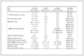
结果1.临床特征:≥2级RP患者在训练组中占38.0%(38/100),在内部验证组中占30.2%(13/43),在外部验证组中占32.4%(11/34)。≥2级RP组与<2级RP组在年龄、性别、吸烟、病理、T分期、肺气肿、ECOG-PS、是否接受免疫治疗、总剂量等指标比较差异均无统计学意义(P> 0.05),而<2级RP组的MLD(U=601.5,P<0.001)、V5(t=-2.03,P=0.045)、V20(U=668.5,P<0.001)和V30(t=-5.61,P<0.001)均低于≥2级RP组,组间差异均具有统计学意义(表 1),logistic回归分析结果显示MLD、V20和V30均是临床发生≥2级RP的独立预测因子,用于建立CM。
|
表 1 不同参数在放射性肺炎分组间具有统计学差异的临床特征分析 Table 1 Analysis of clinical features with significant statistical differences among groups of radiation pneumonitis |
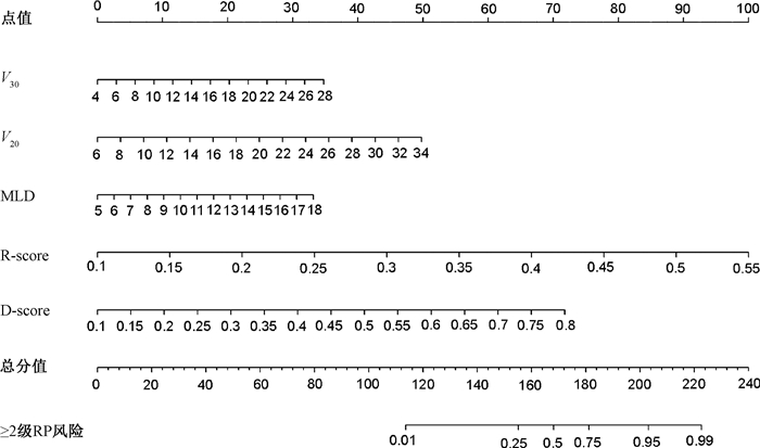
2.特征筛选及模型建立:本研究在训练组(n=100)中进行了特征筛选,最终筛选出14个影像组学特征和15个剂量组学特征,采用MLP机器学习方法分别建立RM、DM,其中影像组学特征中包含5个经过小波滤波器处理的特征,剂量组学特征中包含6个经过小波滤波器处理的特征。利用加权系数分别计算出训练组、内部验证组及外部验证组的R-score和D-score(表 2)。使用PyTorch 1.8.0软件,基于临床独立预测因子(MLD、V20、V30)和R-score、D-score对模型进行可视化,构建预测RP的联合模型(RDN)的列线图,见图 1。
|
|
表 2 各组中不同放射性肺炎分级的R-score与D-score比较 Table 2 Comparison of R-score and D-score of varying RP grades across various groups |
|
注:R-score.影像组学分值;D-score.剂量组学分值;MLD.肺平均剂量;V30.接受≥30 Gy剂量的体积比例;V20.接受≥20 Gy剂量的体积比例;RP. 放射性肺炎 图 1 基于训练组,结合临床特征、R-score、D-score构建联合模型(RDN)的列线图 Figure 1 Nomogram of the RDN constructed by integrating clinical features, R-score, and D-score based on the training group |
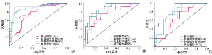
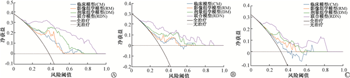
3.模型性能评估:CM、RM、DM和RDN在训练组、内部验证组及外部验证组中的AUC、准确度、灵敏度和特异度见表 3。RDN在训练组、内部验证组及外部验证组中均表现出较好的≥2级RP的预测效果,AUC分别为0.915(95%CI:0.852~0.978)、0.879(95%CI:0.777~0.982)、0.838(95%CI:0.701~ 0.975)(图 2),均显著高于CM(Z=3.76、2.99、2.564,P < 0.05)。DCA显示,在大多数合理阈值概率范围内,RDN比其他模型具有更高的总体净收益(图 3)。
|
|
表 3 各组间不同模型对放射性肺炎的预测性能比较 Table 3 Comparison of performance of different models for RP prediction across groups |
|
图 2 各组不同模型的受试者工作特征曲线 A.训练组;B.内部验证组;C.外部验证组 Figure 2 Receiver operating characteristic (ROC) curves of different models across groups A. Training group, B. Internal validation group, C. External validation group |
|
图 3 各组不同模型的决策曲线(DCA) A.训练组;B.内部验证组;C.外部验证组 Figure 3 Decision curve analysis (DCA) of different models across groups A. Training group; B. Internal validation group; C. External validation group |
讨论
放射性肺炎是接受胸部放疗的非小细胞肺癌患者的重要不良事件,主要通过糖皮质激素治疗,严重者可影响患者的生活质量,甚至导致死亡,因此,在放射治疗前识别放射性肺炎的高危患者并采取预防措施至关重要[4, 9-10]。既往研究表明,一些临床因素,如肺功能、吸烟史、肿瘤部位、肺间质疾病、肺气肿、免疫治疗等与RP的发生相关[4, 11-12]。吸烟和放射性肺炎之间的关系是复杂的。一些研究认为,由于吸烟引起的缺氧和免疫抑制作用,肺的辐射耐受性可能会增加[11, 13]。而本研究中,吸烟在≥2级RP组及<2级RP组中差异无统计学意义,推测可能与本研究未对吸烟状态详细划分有关,比如部分患者既往有吸烟史,但已戒烟多年,本研究仍将患者纳入吸烟队列中,可能会对结果有一定影响。RP的发病率和严重程度与肺内剂量分布有直接关系。剂量学参数如MLD、V5、V10、V20、V30被用于预测RP[4, 7, 12, 14]。本研究中,MLD、V20和V30是≥2级RP发生的独立预测因子,但其预测能力一般,训练组、内部验证组和外部验证组的AUC分别为0.750、0.744、0.660,预测性能仍有待提高。Liang等[15-16] 的研究表明,同侧肺局部剂量变化越大,全肺低剂量区越大,RP发生的概率越大。然而,DVH参数中仅能体现靶体内的二维剂量分布,不能反映局部剂量变化、空间剂量分布或器官结构[8]。
随着影像组学广泛应用于医学研究中,影像组学通过从医学图像中提取大量的高通量特征来改善肿瘤的诊断、预后和治疗决策支持。Krafft等[17] 利用全肺CT影像组学特征预测≥3级RP的AUC为0.68,高于仅使用临床和剂量学参数的预测性能。孔燕等[18] 结合影像组学特征、剂量学参数预测≥2级RP的AUC为0.757。Jiang等[19] 利用定位CT的多个感兴趣区(ROI)内的剂量学因子和影像组学特征建立机器学习模型,结果显示,以全肺-PTV (TL-PTV)作为ROI建立的模型在预测症状性RP方面优于TL-GTV、PTV和GTV等。本研究以TL-PTV作为ROI,利用影像组学特征预测≥2级RP的AUC在训练组、内部验证组和外部验证组中的预测效能均高于DVH参数。剂量组学可以描述靶体内的三维剂量分布,反映出剂量的空间异质性[8, 20]。在Liang等[15] 的研究中,剂量组学可以有效提取剂量分布的空间特征,预测RP的AUC分别为0.709和0.782,均高于DVH参数的AUC(0.665和0.676)。本研究利用剂量组学预测≥2级RP的AUC在训练组、内部验证组和外部验证组亦高于DVH参数。既往大部分研究仅使用影像组学或剂量组学单一组学进行分析,本研究联合了影像组学、剂量组学及剂量学参数进行多维数据综合分析,其联合模型的预测性能最佳,在训练组、内部验证组和外部验证组的AUC结果表明,该预测模型具有良好的泛化能力、稳定性及实用性。此外本研究在提取影像组学、剂量组学特征之前,进行了滤波处理,去除噪声影响,并重新计算纹理特征和一阶特征,以描述病变不同频段的特征,提取的特征更能反映组织成像的本质内容[18, 21]。
本研究仍有一定局限性。首先,作为回顾性研究,样本量较小;其次,在对本研究病例随访过程中,发现有23例患者放疗后失访,未纳入研究中,也可能对结果产生一定影响;另外,免疫治疗和一些化疗药物对RP有影响[7, 22],本研究未对其深入研究。下一步将纳入前瞻性研究,严格控制患者纳入标准,降低失访率。
综上所述,本研究发现,基于定位CT和剂量学图像的影像组学、剂量组学和剂量学参数建立的联合模型可有效预测≥2级RP的发生,可实现在放疗前个体化预测≥2级RP的发生,对临床早期干预和治疗实施具有重要价值,从而使NSCLC放疗患者受益。
利益冲突 无
作者贡献声明 王寻、卞婷婷负责实验设计、研究实施和论文撰写;丁强、张爱平、韩心舒、张谷青负责收集、处理患者资料;葛双、马俊丽协助实验研究、统计分析和论文修改;陈月芹、叶书成指导论文修改
| [1] |
Jia B, Zheng Q, Wang J, et al. A nomogram model to predict death rate among non-small cell lung cancer (NSCLC) patients with surgery in surveillance, epidemiology, and end results (SEER) database[J]. BMC Cancer, 2020, 20(1): 666. DOI:10.1186/s12885-020-07147-y |
| [2] |
Siegel RL, Miller KD, Wagle NS, et al. Cancer statistics, 2023[J]. CA Cancer J Clin, 2023, 73(1): 17-48. DOI:10.3322/caac.21763 |
| [3] |
Arroyo-Hernández M, Maldonado F, Lozano-Ruiz F, et al. Radiation-induced lung injury: current evidence[J]. BMC Pulm Med, 2021, 21(1): 9. DOI:10.1186/s12890-020-01376-4 |
| [4] |
Yang S, Huang S, Ye X, et al. Risk analysis of grade ≥ 2 radiation pneumonitis based on radiotherapy timeline in stage Ⅲ/Ⅳ non-small cell lung cancer treated with volumetric modulated arc therapy: a retrospective study[J]. BMC Pulm Med, 2022, 22(1): 402. DOI:10.1186/s12890-022-02211-8 |
| [5] |
Chen M, Copley SJ, Viola P, et al. Radiomics and artificial intelligence for precision medicine in lung cancer treatment[J]. Semin Cancer Biol, 2023, 93: 97-113. DOI:10.1016/j.semcancer.2023.05.004 |
| [6] |
Tunali I, Gillies RJ, Schabath MB. Application of radiomics and artificial intelligence for lung cancer precision medicine[J]. Cold Spring Harb Perspect Med, 2021, 11(8): a039537. DOI:10.1101/cshperspect.a039537 |
| [7] |
Zhang Z, Wang Z, Yan M, et al. Radiomics and dosiomics signature from whole lung predicts radiation pneumonitis: A model development study with prospective external validation and decision-curve analysis[J]. Int J Radiat Oncol Biol Phys, 2023, 115(3): 746-758. DOI:10.1016/j.ijrobp.2022.08.047 |
| [8] |
Huang Y, Feng A, Lin Y, et al. Radiation pneumonitis prediction after stereotactic body radiation therapy based on 3D dose distribution: dosiomics and/or deep learning-based radiomics features[J]. Radiat Oncol, 2022, 17(1): 188. DOI:10.1186/s13014-022-02154-8 |
| [9] |
闫萌, 张臻, 于佳琦, 等. 基于剂量组学预测肺癌患者放射性肺炎发生的研究[J]. 中华放射肿瘤学杂志, 2022, 31(8): 698-703. Yan M, Zhang Z, Yu JQ, et al. Dosiomics-based prediction of incidence of radiation pneumonitis in lung cancer patients[J]. Chin J Radiat Oncol, 2022, 31(8): 698-703. DOI:10.3760/cma.j.cn113030-20211115-00466 |
| [10] |
Zha Y, Zhang J, Yan X, et al. A dynamic nomogram predicting symptomatic pneumonia in patients with lung cancer receiving thoracic radiation[J]. BMC Pulm Med, 2024, 24(1): 99. DOI:10.1186/s12890-024-02899-w |
| [11] |
Zhang XJ, Sun JG, Sun J, et al. Prediction of radiation pneumonitis in lung cancer patients: a systematic review[J]. J Cancer Res Clin Oncol, 2012, 138(12): 2103-2116. DOI:10.1007/s00432-012-1284-1 |
| [12] |
Palma DA, Senan S, Tsujino K, et al. Predicting radiation pneumonitis after chemoradiation therapy for lung cancer: an international individual patient data meta-analysis[J]. Int J Radiat Oncol Biol Phys, 2013, 85(2): 444-450. DOI:10.1016/j.ijrobp.2012.04.043 |
| [13] |
Vogelius IR, Bentzen SM. A literature-based meta-analysis of clinical risk factors for development of radiation induced pneumonitis[J]. Acta Oncol, 2012, 51(8): 975-983. DOI:10.3109/0284186X.2012.718093 |
| [14] |
王谨, 包勇, 庄婷婷, 等. 非小细胞肺癌同期放化疗后重度急性放射性肺炎的预测模型研究[J]. 中华放射肿瘤学杂志, 2013, 22(6): 455-459. Wang J, Bao Y, Zhuang TT, et al. Study on prediction models for severe acute radiation pneumonitis in patients with non-small cell lung cancer after concurrent chemoradiotherapy[J]. Chin J Radiat Oncol, 2013, 22(6): 455-459. DOI:10.3760/cma.j.issn.1004-4221.2013.06.009 |
| [15] |
Liang B, Yan H, Tian Y, et al. Dosiomics: extracting 3D spatial features from dose distribution to predict incidence of radiation pneumonitis[J]. Front Oncol, 2019, 9: 269. DOI:10.3389/fonc.2019.00269 |
| [16] |
Liang B, Tian Y, Chen X, et al. Prediction of radiation pneumonitis with dose distribution: A convolutional neural network (CNN) based model[J]. Front Oncol, 2019, 9: 1500. DOI:10.3389/fonc.2019.01500 |
| [17] |
Krafft SP, Rao A, Stingo F, et al. The utility of quantitative CT radiomics features for improved prediction of radiation pneumonitis[J]. Med Phys, 2018, 45(11): 5317-5324. DOI:10.1002/mp.13150 |
| [18] |
孔燕, 吴佳, 魏贤顶, 等. 肺癌放疗患者症状性放射性肺炎预测的CT影像组学研究[J]. 中华放射医学与防护杂志, 2022, 42(2): 115-120. Kong Y, Wu J, Wei XD, et al. Application of CT radiomics analysis to predict symptomatic radiation pneumonitis for lung cancer[J]. Chin J Radiol Med Prot, 2022, 42(2): 115-120. DOI:10.3760/cma.j.cn112271-20210730-00301 |
| [19] |
Jiang W, Song Y, Sun Z, et al. Dosimetric factors and radiomics features within different regions of interest in planning CT images for improving the prediction of radiation pneumonitis[J]. Int J Radiat Oncol Biol Phys, 2021, 110(4): 1161-1170. DOI:10.1016/j.ijrobp.2021.01.049 |
| [20] |
Mylona E, Acosta O, Lizee T, et al. Voxel-based analysis for identification of urethrovesical subregions predicting urinary toxicity after prostate cancer radiation therapy[J]. Int J Radiat Oncol Biol Phys, 2019, 104(2): 343-354. DOI:10.1016/j.ijrobp.2019.01.088 |
| [21] |
Jing R, Wang J, Li J, et al. A wavelet features derived radiomics nomogram for prediction of malignant and benign early-stage lung nodules[J]. Sci Rep, 2021, 11(1): 22330. DOI:10.1038/s41598-021-01470-5 |
| [22] |
Chen F, Niu J, Wang M, et al. Re-evaluating the risk factors for radiation pneumonitis in the era of immunotherapy[J]. J Transl Med, 2023, 21(1): 368. DOI:10.1186/s12967-023-04212-5 |