 2025, Vol. 45


2. 山西省肿瘤医院 中国医学科学院肿瘤医院山西医院 山西医科大学附属肿瘤医院放射治疗科,太原 030013
2. Department of Radiotherapy, Shanxi Province Cancer Hospital/Shanxi Hospital Affiliated to Cancer Hospital, Chinese Academy of Medical Sciences/Cancer Hospital Affiliated to Shanxi Medical University, Taiyuan 030013, China
食管癌(EC)在全球常见癌症中居第11位,同时在癌症相关死亡中居第7位[1]。超过一半的食管癌患者在确诊时为不可切除局部晚期或转移性疾病[2]。非免疫治疗时代,治疗以化疗为主,但疗效差,中位生存期仅为9.5~13.1个月[3-4]。近年来多项Ⅲ期临床研究结果显示,免疫联合化疗使得晚期食管癌预后有所提升,中位生存期提高到15.3~20.0个月,并已经确立其为晚期食管癌的标准治疗。但无进展生存期仅为6.9~10个月,预后还有很大的提升空间[5-6]。
放射治疗是EC的主要治疗手段之一,对于手术不耐受或手术难度大的早期EC患者,放疗是可选择的替代治疗方法;对于可手术/不可切除的局部晚期EC患者,分别以术前新辅助放化疗和根治性放化疗作为标准治疗方案;对于晚期EC患者,放疗联合化疗可以为寡转移患者带来生存获益,提高患者生活质量[7]。那么,化免基础上联合放疗是否能进一步改善晚期EC患者预后,目前鲜有证据支持。为此,本研究旨在探讨一线免疫化疗后联合放疗治疗晚期食管癌疗效。
资料与方法1.患者一般资料:回顾2018年1月至2023年5月在山西省肿瘤医院连续接受治疗的食管癌患者资料。入组标准:病理确诊为鳞癌;按美国癌症联合会(American Joint Committee on Cancer,AJCC)第8版分期,ⅣA/ⅣB期;接受一线化免联合或不联合放射治疗。排除标准:原发性食管肿瘤和区域淋巴结的放疗剂量 < 30 Gy;一线化免前已接受抗肿瘤治疗;病历资料不完整;随访时间<90 d。
治疗前检查包括血液学和生化检查、食管胃十二指肠镜活检、胸部CT、腹部CT、颈部彩超、上消化道钡餐,部分患者必要时行正电子发射计算机体层成像(PET)-CT或胸部磁共振成像(MRI)。
本研究经山西医科大学附属肿瘤医院/中国医学科学院肿瘤医院/山西省肿瘤医院伦理委员会批准(伦理编号:2024-122),免除知情同意。
2.治疗:根据是否接受放射治疗,分为化免联合放疗组和化免组。化免联合放疗组患者接受一线免疫化疗,同步或随后接受放疗,伴或不伴巩固免疫治疗。化免组患者仅接受一线免疫化疗。患者平均接受4个周期化疗免疫治疗。常用的免疫检查点抑制剂包括卡瑞丽珠单抗、替雷利珠单抗、特瑞普利单抗、帕博丽珠单抗、信迪利单抗、纳武利尤单抗。最常用的诱导和同步化疗方案是紫杉醇类联合铂类。一部分患者由于年龄较大或化疗的不良事件而选择口服S-1作为化疗方案。采用调强放射治疗(IMRT),能量为6 MV X射线。原发肿瘤的大体肿瘤体积(GTVp)定义为原发性食管肿瘤,淋巴结的大体肿瘤体积(GTVnd)定义为CT或PET/CT扫描或细胞学证实的阳性淋巴结。临床靶体积(CTV)定义为GTVp加上近端和远端边缘3 cm, GTVnd加上0.5~1.0 cm边缘。根据原发肿瘤的位置,还包括预防性区域淋巴引流区域。计划大体肿瘤体积(PGTV)和计划靶体积(PTV)分别在三维方向上超出GTV或CTV 5 mm。对于有远处转移的患者,一般将颈、腹腔阳性淋巴结纳入照射野,肺、肝、骨等远处器官可选择局部治疗。PGTV和PTV的处方剂量分别为60和50 Gy,采用同步加量(simultaneous integrated boost, SIB)技术,每周连续5 d,共28~30次。
患者在前两年每3个月随访一次,在接下来的两年中每6个月随访一次,5年后每年随访一次。临床检查包括常规血液检查和肿瘤标志物、胸部CT/增强CT、颈部/腹部CT或超声、钡餐。通过影像学或组织细胞学活检证实复发。局部淋巴结复发(LRR)定义为原发肿瘤或区域淋巴结的任何复发。非区域淋巴结或远处部位的进展被认为是远处转移(DM)。
3.统计学处理:采用R软件4.4.2版本进行分析。分类变量组间比较,当理论频数≥5时采用χ2检验,当理论频数 < 5时采用Fisher精确检验。倾向性评分加权(IPTW)用于调整不平衡的混杂因素。总生存期(overall survival,OS)计算从治疗开始之日起至死亡或随访截止时间。无进展生存期(progression free survival,PFS)计算从治疗开始之日起至复发、死亡或随访截止时间。使用Kaplan-Meier方法绘制生存曲线,并通过对数秩检验进行比较。使用ggadjustedcurves函数(survminer包0.4.9版本)通过Cox风险比例模型对生存曲线进行校正。单变量分析采用Cox比例风险模型。P < 0.05为差异有统计学意义。
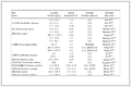
结果1.基线特征:本研究共纳入137例Ⅳ期食管鳞癌患者,其中化免联合放疗组43例(31.4%),单纯化免组94例(68.6%)。联合放疗组与单纯化免组中≥65岁患者比例相近(53.5% vs 52.1%),男性占比分别为60.5%和71.3%。联合放疗组T4期患者比例较高(30.2% vs 22.3%),但M1期患者比例较低(60.5% vs 69.1%)。两组N分期分布相似,N3期比例分别为18.6%和22.3%。ECOG 0~1分患者占大多数(联合组76.7%,化免组86.2%),营养风险评分(NRS≥3)比例分别为23.3%和28.7%。通过逆概率治疗加权(IPTW)校正后,所有变量的标准化均数差(SMD)均 < 0.1(表 1)。
|
表 1 IPTW前后不同治疗模式组晚期食管鳞癌患者临床病理特征比较[n(%)] Table 1 Comparison of clinicopathologic features of patients with advanced ESCC treated using different strategies before and after calibration using IPTW [n(%)] |
2.预后影响因素分析:将可能影响患者预后的因素如治疗模式、年龄、性别、临床分期、病灶位置、吸烟史、东部肿瘤协作组(ECOG)体能状态评分以及NRS-2002评分纳入Cox单因素回归模型进行分析,结果显示,仅有放疗与患者的PFS和OS相关(P<0.05),其余因素均未发现与患者预后显著相关(P>0.05,表 2)。
|
|
表 2 影响晚期食管鳞癌患者PFS和OS的单因素Cox回归分析 Table 2 Univariate Cox regression analysis of factors influencing PFS and OS of patients with advanced ESCC |
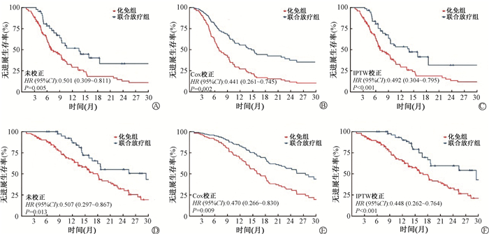
3.生存分析:由图 1可见患者预后。化免联合放疗组的中位PFS、中位OS均显著优于化免组。校正前化免组中位PFS为7.0个月,化免联合放疗组为13.6个月(HR=0.501,95%CI:0.309~0.811,P = 0.005);Cox校正年龄、性别、ECOG PS评分、吸烟史、T分期、N分期、M分期、肿瘤位置后化免组中位PFS为7.0个月,化免联合放疗组为14.7个月(HR=0.441,95% CI:0.261~0.745,P = 0.002);IPTW后(标准化平均差均 < 10%),化免组中位PFS为7.0月,化免联合放疗组为13.9月(HR=0.492,95% CI:0.304~0.795, P < 0.001)。校正前化免组中位OS分别为18.0月,化免联合放疗组为29.5月(HR=0.507,95%CI:0.297~0.867,P = 0.013)。Cox校正年龄、性别、ECOG PS评分、吸烟史、T分期、N分期、M分期、肿瘤位置后化免组中位OS分别为16.7月,化免联合放疗组为27.5月(HR=0.470, 95% CI:0.266~0.830, P = 0.009)。IPTW后化免组中位OS分别为16.9个月,化免联合放疗组为29.5个月(HR=0.448, 95% CI: 0.262~ 0.764,P < 0.001)。
|
图 1 采用不同方案治疗晚期食管鳞癌患者的生存曲线比较 A.校正前无进展生存;B.Cox校正后无进展生存;C.IPTW后无进展生存;D.校正前总生存;E.Cox校正后总生存;F.IPTW后总生存 Figure 1 Comparison of survival curves for patients with advanced ESCC treated using varying therapies A. Pre-calibration PFS; B. PFS after calibration using the COX model; C. PFS after calibration using IPTW; D. Pre-calibration overall survival; E. Overall survival after calibration using the Cox model; F. Overall survival after calibration using IPTW |
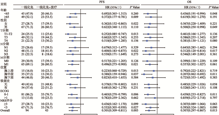
4.亚组分析:对OS和PFS进行亚组分析,以探讨一线化疗免疫治疗后放疗的潜在益处。如图 2所示,在按年龄、性别、ECOG评分、吸烟史、T分期、N分期、M分期及肿瘤位置定义的绝大多数亚组中,相比一线化疗免疫,化免联合放疗在OS和PFS中均有获益趋势。
|
图 2 化免和化免联合放疗在不同人群和临床特征下的无进展生存期(PFS)和总生存期(OS)的比较 Figure 2 Comparison of PFS and OS under different populations and clinical features between sole chemoimmunotherapy and chemoimmunotherapy + radiotherapy |
5.患者的治疗相关不良反应:由表 3可见,放射治疗可能会增加食管炎、≥3级骨髓抑制和1~2级肺炎风险。最常见的不良反应是消化道反应和骨髓抑制。化免联合放疗组与单纯化学免疫组的1~2级消化道反应(46.5% vs. 40.4%,P = 0.504)、≥3级消化道反应(16.3% vs. 21.3%,P = 0.495)、1~2级骨髓抑制(44.2% vs. 51.1%,P = 0.455)、1~2级肝损伤(2.3% vs. 5.3%,P = 0.665)、≥3级肝损伤(0.0% vs. 2.2%,P = 0.999)和≥3级放射性肺炎(2.3% vs 0%,P = 0.314)发生率上差异均无统计学意义(P>0.05)。化免联合放疗组≥3级骨髓抑制的发生率显著高于单纯化免组(30.2% vs. 13.1%,χ2=7.011, P = 0.008)。此外,联合放疗显著增加了1~2级食管炎(23.3% vs. 1.1%, P < 0.001)和1~2级肺炎(37.2% vs. 2.1%, P < 0.001)的风险。在≥3级食管炎方面,仅联合放疗组观察到5例(11.6%),单纯化免组为0例(P = 0.003)。未观察到治疗相关死亡病例。
|
|
表 3 不同治疗模式晚期食管鳞癌患者治疗相关不良反应比较 Table 3 Comparison of treatment-related adverse reactions among patients with advanced ESCC treated using different strategies |
讨论
已有一些研究阐明了放疗联合化疗在晚期ESCC中的价值。一项回顾性分析,比较了接受化疗或联合放疗的IV期ESCC患者,发现放疗可以延长OS,从11个月延长到14个月[8]。然而,Chen等[7] 发现联合放射治疗不能延长少转移鳞癌患者的中位OS (16.8个月vs. 14.8个月,P = 0.056)。联合放疗和化学免疫治疗治疗晚期食管鳞癌的可行性和临床益处仍不确定。本研究表明化免联合放疗具有显著的生存优势。
本研究采用Cox比例风险模型校正年龄、性别及临床病理特征后,化免联合放疗组的风险比进一步降低,提示协变量调整可能更精准地揭示了放疗联合治疗的独立生存获益。这一变化可能与基线特征对治疗效应的潜在混杂作用有关,例如,若放疗组中合并更多高龄或晚期(如T/N分期较高)患者,未校正模型可能低估其真实疗效;校正后模型则可排除此类偏倚,从而凸显治疗本身的效应。由于回顾性研究的内在局限性和选择偏差的可能,我们进行了IPTW分析,以更准确地观察放射治疗的效果。IPTW分析显示,与单纯免疫化疗相比,放疗在PFS和OS中仍显示出优势。这项研究阐明了将放疗与免疫化疗结合起来作为晚期食管鳞癌的一线治疗方法的有效性。
放疗通过激活抗肿瘤免疫应答发挥协同作用:促进抗原释放与呈递[9-11]、增加淋巴细胞浸润[12-13]、重塑肿瘤微环境[14-15],为放疗联合免疫治疗奠定基础[16-17]。在食管癌领域,多项临床研究验证了联合治疗模式的潜力。TENERGY研究的长期随访数据显示,同步放化疗后序贯阿替利珠单抗维持治疗的中位OS达31.0个月,显示出良好的耐受性[18]。对于转移性/难治性患者,卡瑞利珠单抗联合同步放疗(30~50 Gy/10~25次)的Ⅱ期研究显示,中位PFS和OS分别为6.9和12.8个月,3~4级不良事件发生率控制在63.3%[19]。值得注意的是,Wu等[20] 认为放疗介入时机可能影响疗效,免疫治疗前后90 d内接受放疗者较延迟放疗组生存显著改善(HR=0.73,P=0.04),提示放疗应作为初始治疗策略而非疾病进展后的补救措施。现有证据表明,免疫治疗联合放疗在晚期食管鳞癌中展现出有前景的疗效和可控的安全性,但需更多Ⅲ期随机对照试验验证最佳联合模式。未来研究应重点关注放射剂量分割方案、治疗时序安排以及生物标志物指导下的个体化治疗策略。
本研究结果表明,免疫治疗联合放疗虽可增加1~2级放射性肺炎发生率,但未显著升高≥3级放射性肺炎风险。值得注意的是,联合治疗组≥3级食管炎及骨髓抑制发生率较单纯免疫治疗有所增加,但未观察到治疗相关死亡病例。综合现有证据提示,免疫治疗联合放疗用于晚期食管癌患者具有可控的安全性特征,其毒性谱虽存在特征性差异,但整体耐受性良好。
食管癌是一种侵袭性疾病,通常在晚期被诊断。90%以上的患者以吞咽困难为首发症状[21]。食管癌患者更容易营养不良,这显著降低了患者的预后[22-23]。对于食管癌患者,PNI、PG-SGA、NRS-2002等工具是较常用的评估方法[24]。已有研究表明NRS-2002营养风险评分增加与生存期(OS和PFS)降低相关[25]。然而在免疫治疗时代下,NRS与患者预后的关系仍需进一步研究,在本研究中未发现NRS评分与患者OS、PFS显著相关。
本研究存在的局限:首先,作为回顾性研究,尽管通过逆概率加权(IPTW)调整了基线混杂因素,但回顾性研究无法完全控制未测量的混杂变量(如分子标志物表达、合并症细节),可能影响结果的因果推断;其次,总样本量较小,尤其在亚组分析中统计效能可能不足,导致部分潜在差异未被检出(如分期或肿瘤位置的亚组);此外,数据来自单中心,缺乏外部数据验证。
对于无法切除的局部晚期或转移性食管癌患者,一线化疗免疫联合放疗可有效改善局部控制和生存结果。需要进一步的前瞻性证据来验证这些结果。
利益冲突 无
作者贡献声明 丰培鑫负责数据整理和论文撰写;侯庆、姚宁宁负责研究设计;张文娟、孙博辰、牛文霞、赵岸琪、陈文路、吴白雪、周钰滢、张译文负责数据整理;梁玉、曹欣、柏玮负责统计分析;刘建庭、张双平、曹建忠指导论文修改
| [1] |
郑渊杰, 滕熠, 何思怡, 等. 2022年全球及中国食管癌流行病学特征[J]. 中国肿瘤, 2025, 34(3): 165-170. Zheng YJ, Teng Y, He SY, et al. Epidemiological characteristics of esophageal cancer worldwide and in China, 2022[J]. China Cancer, 2025, 34(3): 165-170. DOI:10.11735/j.issn.1004-0242.2025.03.A001 |
| [2] |
van Rossum P, Mohammad NH, Vleggaar FP, et al. Treatment for unresectable or metastatic oesophageal cancer: current evidence and trends[J]. Nat Rev Gastroenterol Hepatol, 2018, 15(4): 235-249. DOI:10.1038/nrgastro.2017.162 |
| [3] |
Lorenzen S, Schuster T, Porschen R, et al. Cetuximab plus cisplatin-5-fluorouracil versus cisplatin-5-fluorouracil alone in first-line metastatic squamous cell carcinoma of the esophagus: a randomized phase Ⅱ study of the Arbeitsgemeinschaft Internistische Onkologie[J]. Ann Oncol, 2009, 20(10): 1667-1673. DOI:10.1093/annonc/mdp069 |
| [4] |
Shinoda M, Ando N, Kato K, et al. Randomized study of low-dose versus standard-dose chemoradiotherapy for unresectable esophageal squamous cell carcinoma (JCOG0303)[J]. Cancer Sci, 2015, 106(4): 407-412. DOI:10.1111/cas.12622 |
| [5] |
Luo H, Lu J, Bai Y, et al. Effect of camrelizumab vs placebo added to chemotherapy on survival and progression-free survival in patients with advanced or metastatic esophageal squamous cell carcinoma: The ESCORT-1st randomized clinical trial[J]. JAMA, 2021, 326(10): 916-925. DOI:10.1001/jama.2021.12836 |
| [6] |
Janjigian YY, Kawazoe A, Bai Y, et al. Pembrolizumab plus trastuzumab and chemotherapy for HER2-positive gastric or gastro-oesophageal junction adenocarcinoma: interim analyses from the phase 3 KEYNOTE-811 randomised placebo-controlled trial[J]. Lancet, 2023, 402(10418): 2197-2208. DOI:10.1016/S0140-6736(23)02033-0 |
| [7] |
Chen Y, Cheng X, Song H, et al. Outcomes of concurrent chemoradiotherapy versus chemotherapy alone for esophageal squamous cell cancer patients presenting with oligometastases[J]. J Thorac Dis, 2019, 11(4): 1536-1545. DOI:10.21037/jtd.2019.03.10 |
| [8] |
Lyu J, Li T, Wang Q, et al. Outcomes of concurrent chemoradiotherapy versus chemotherapy alone for stage Ⅳ esophageal squamous cell carcinoma: a retrospective controlled study[J]. Radiat Oncol, 2018, 13(1): 233. DOI:10.1186/s13014-018-1183-y |
| [9] |
Lin W, Xu Y, Chen X, et al. Radiation-induced small extracellular vesicles as " carriages" promote tumor antigen release and trigger antitumor immunity[J]. Theranostics, 2020, 10(11): 4871-4884. DOI:10.7150/thno.43539 |
| [10] |
Sharabi AB, Nirschl CJ, Kochel CM, et al. Stereotactic radiation therapy augments antigen-specific PD-1-mediated antitumor immune responses via cross-presentation of tumor antigen[J]. Cancer Immunol Res, 2015, 3(4): 345-355. DOI:10.1158/2326-6066.CIR-14-0196 |
| [11] |
Zhou Z, Zhao J, Hu K, et al. Single high-dose radiation enhances dendritic cell homing and T cell priming by promoting Reactive Oxygen Species-induced cytoskeletal reorganization[J]. Int J Radiat Oncol Biol Phys, 2021, 109(1): 95-108. DOI:10.1016/j.ijrobp.2020.07.2321 |
| [12] |
Anitei MG, Zeitoun G, Mlecnik B, et al. Prognostic and predictive values of the immunoscore in patients with rectal cancer[J]. Clin Cancer Res, 2014, 20(7): 1891-1899. DOI:10.1158/1078-0432.CCR-13-2830 |
| [13] |
Kovács A, Stenmark Tullberg A, Werner Rönnerman E, et al. Effect of radiotherapy after breast-conserving surgery depending on the presence of tumor-infiltrating lymphocytes: a long-term follow-up of the SweBCG91RT randomized trial[J]. J Clin Oncol, 2019, 37(14): 1179-1187. DOI:10.1200/JCO.18.02157 |
| [14] |
Barker HE, Paget JT, Khan AA, et al. The tumour microenvironment after radiotherapy: mechanisms of resistance and recurrence[J]. Nat Rev Cancer, 2015, 15(7): 409-425. DOI:10.1038/nrc3958 |
| [15] |
McLaughlin M, Patin EC, Pedersen M, et al. Inflammatory microenvironment remodelling by tumour cells after radiotherapy[J]. Nat Rev Cancer, 2020, 20(4): 203-217. DOI:10.1038/s41568-020-0246-1 |
| [16] |
Demaria S, Coleman CN, Formenti SC. Radiotherapy: changing the game in immunotherapy[J]. Trends Cancer, 2016, 2(6): 286-294. DOI:10.1016/j.trecan.2016.05.002 |
| [17] |
Jarosz-Biej M, Smolarczyk R, Cichoń T, et al. Tumor microenvironment as a "game changer" in cancer radiotherapy[J]. Int J Mol Sci, 2019, 20(13): 3212. DOI:10.3390/ijms20133212 |
| [18] |
Bando H, Kumagai S, Kotani D, et al. Atezolizumab following definitive chemoradiotherapy in patients with unresectable locally advanced esophageal squamous cell carcinoma - a multicenter phase 2 trial (EPOC1802)[J]. Nat Cancer, 2025, 6(3): 445-459. DOI:10.1038/s43018-025-00918-1 |
| [19] |
Zhao W, Ke S, Cai X, et al. Radiotherapy plus camrelizumab and irinotecan for oligometastatic esophageal squamous cell carcinoma patients after first-line immunotherapy plus chemotherapy failure: An open-label, single-arm, phase Ⅱ trial[J]. Radiother Oncol, 2023, 184: 109679. DOI:10.1016/j.radonc.2023.109679 |
| [20] |
Wu X, Li Y, Zhang K, et al. Immunotherapy with or without radiotherapy for metastatic or recurrent esophageal squamous cell carcinoma: A real-world study[J]. Clin Transl Radiat Oncol, 2023, 38: 130-137. DOI:10.1016/j.ctro.2022.10.011 |
| [21] |
Moses FM. Squamous cell carcinoma of the esophagus. Natural history, incidence, etiology, and complications[J]. Gastroenterol Clin North Am, 1991, 20(4): 703-716. DOI:10.1007/BF02050082 |
| [22] |
Cao J, Xu H, Li W, et al. Nutritional assessment and risk factors associated to malnutrition in patients with esophageal cancer[J]. Curr Probl Cancer, 2021, 45(1): 100638. DOI:10.1016/j.currproblcancer.2020.100638 |
| [23] |
Antasouras G, Papadopoulou SK, Tolia M, et al. May nutritional status positively affect disease progression and prognosis in patients with esophageal and pharyngeal cancers? A scoping review of the current clinical studies[J]. Med Sci (Basel), 2023, 11(4): 64. DOI:10.3390/medsci11040064 |
| [24] |
于玲, 赵佳佳, 姜桂春. 老年食管癌患者营养评估工具的选择和应用[J]. 国际老年医学杂志, 2022, 43(4): 434-437. Yu L, Zhao JJ, Jiang GC. Selection and application of nutritional assessment tools for older patients with esophageal cancer[J]. Int J Geriatr, 2022, 43(4): 434-437. DOI:10.3969/j.issn.1674-7593.2022.04.012 |
| [25] |
Song T, Wan Q, Yu W, et al. Pretreatment nutritional risk scores and performance status are prognostic factors in esophageal cancer patients treated with definitive chemoradiotherapy[J]. Oncotarget, 2017, 8(58): 98974-98984. DOI:10.18632/oncotarget.21940 |