2. 首都医科大学附属北京世纪坛医院 放疗科,北京 100038;
3. 中国疾病预防控制中心辐射防护与核安全医学研究所 中国疾病预防控制中心放射防护与核应急重点实验室 世界卫生组织辐射与健康合作中心,北京 100088
2. Radiotherapy Department, Beijing Shijitan Hospital Affiliated to Capital Medical University, Beijing 100038, China;
3. Key Laboratory of Radiological Protection and Nuclear Emergency, China CDC, WHO Collaborating Centre for Radiation and Health, National Institute for Radiological Protection, Chinese Center for Disease Control and Prevention, Beijijng 100088, China
2020年全球癌症统计报告显示癌症已成为中国人死亡的主要原因,其死亡率跃居全球第一,数据表明,70%的恶性肿瘤在不同阶段需要接受放疗,其中约40%仅通过放疗即可达到根治目的[1]。
螺旋断层放疗(TOMO)系统是一台安装在滑环上的兆伏级(MV)加速器,它采用强度调制的扇形束配合床的运动在360°旋转过程中对患者进行放疗。多篇国内外文献报道其极高的强度调制能力、独特的投照方式对于放疗靶区的剂量分布与危及器官的保护有很大的优势[2-5],但同时也对束流采集与质量控制(QC)带来了挑战[6-8]。TOMO的质量控制对于保证放疗安全性和准确性至关重要[9]。
TOMO治疗计划系统(TPS)的射束模型基于厂家金标准数据建立,用户无法调整此模型。但是,临床使用科室有责任利用各种QC设备测量束流数据从而检验设备运行状态。目前QC所使用的各种设备仪器大多是国外厂商提供,在数据分析上受限,因此自主研发QC设备检测加速器保证放疗的准确性已成为当务之急。本研究应用国产二维水箱测量TOMO束流,并以厂家金标准数据为基准分析差异,进行国产水箱应用于TOMO QC的可行性研究。
材料与方法 1、实验设备本研究中使用型号为Hi-ART的TOMO放疗系统(美国Accuray公司);使用两套水箱系统:水箱1为进口二维水箱TS(美国TomoScanner),水箱自动扫描软件系统为TEMS(Tomotherapy Electrometer Measurement System)2.0版本;水箱2为国产二维水箱,水箱自动扫描软件系统为Engineer V2.0版本。
水箱1和水箱2的特性列于表 1。
|
|
表 1 两个水箱的性能参数 Table 1 Performance parameters of the two water tanks |
2、设备的机械QC
TOMO的束流与机械部分精度密切相关[10]。本研究开始前,根据WS 531-2017《螺旋断层治疗装备质量控制检测规范》[11]对TOMO加速器进行了年检,项目包括:静态输出剂量、旋转输出剂量、射野纵向与横向剂量分布(profile)、百分深度剂量(percentage depth dose,PDD)、多叶准直器(MLC)横向偏移等。在采集束流数据前根据厂家手册再次进行了机械部分QC包括:加速管、钨门(Jaw)、MLC、探测器、治疗床、激光灯,以保证TOMO处于最佳状态。
3、数据采集所有的束流扫描和数据收集严格按照美国医学物理学会(American Association of Physicists in Medicine,AAPM)发布的TG-148[12]、TG-106[13]以及TG-306[14]报告执行。首先将水箱横向放置在平整的床面上对水箱进行初步调平摆位,注入蒸馏水后通过旋转调平点位进行机械臂的调平。将水平面放置在TOMO的等中心处插入电离室,通过兆伏级CT(MVCT)扫描电离室将其放置中间水平位置,再将扫描电离室参考位置下移0.6 r(指型电离室半径的3/5)[15]至有效测量点。将TOMO机架角度固定在0°,MLC全部打开,源轴距(source axis distance,SAD)为85.0 cm,形成40.0 cm×1.0 cm、40.0 cm×2.5 cm、40.0 cm×5.0 cm 3个射野,分别测量水下1.5、5.0、10.0、15.0、20.0 cm深度的横向离轴剂量分布(Profiletrans);然后旋转水箱90°,参照测量横向离轴剂量分布的方法,重新摆位与调平后,形成25.0 cm×1.0 cm、25.0 cm×2.5 cm、25.0 cm×5.0 cm 3个射野,分别测量PDD以及水下1.5、5.0、10.0、15.0、20.0 cm深度的纵向离轴剂量分布(ProfileLong)。
4、数据分析扫描结束后将处理过的数据统一导入到TomoScanner自带的TEMS分析软件中与参考数据进行γ分析,得到相应的四分之一高宽(full width at quarter maximum,FWQM)、半高宽(full width at half maximum,FWHM)和最大γ值(Max γ)。依据设备厂家验收手册,横向离轴剂量分布γ分析标准为2%/1 mm,γ值< 1;测量射野FWQM与参考射野FWQM数据偏差 < 1%。纵向离轴剂量分布γ分析标准为2%/1%射野宽度,γ值< 1;测量射野FWHM与参考射野FWHM数据偏差 < 1%。将水箱2所测数据与金标数据导入GraphPad Prism软件中得到对比曲线,并进行曲线拟合分析。
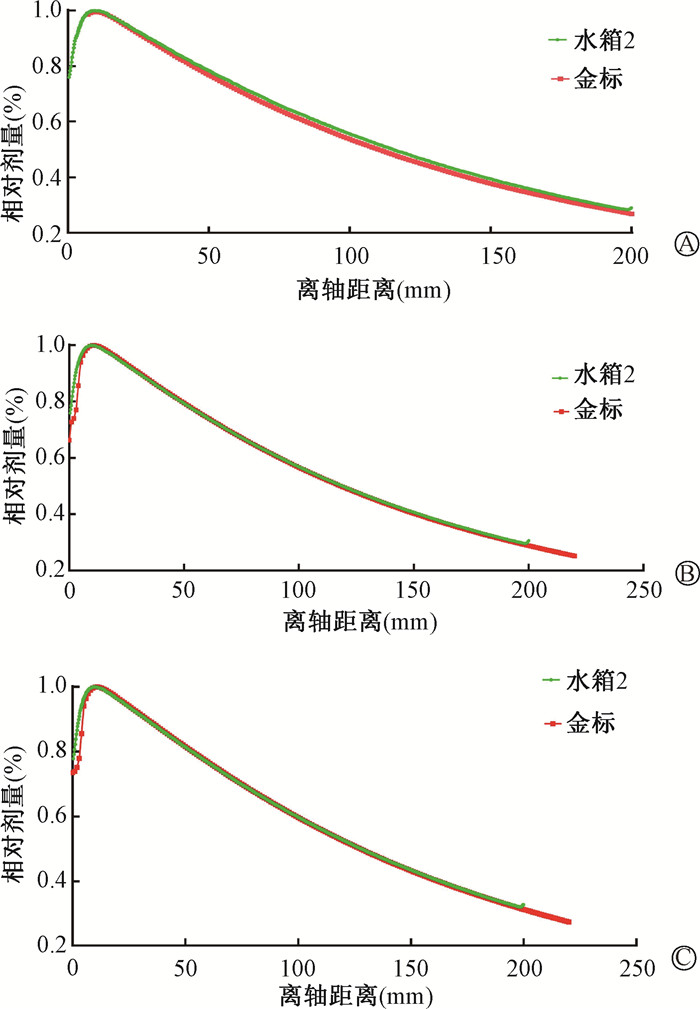
结果 1、PDD结果对3个不同射野下采集的PDD曲线采用最小二乘法进行平滑处理,在最大剂量深度处进行归一。金标和水箱2在3个不同射野下的PDD20/PDD10值均在0.5~0.6之间。水箱2与金标的PDD20/PDD10相对偏差在3个不同射野下均>1%,结果列于表 2。水箱2与金标两者在3个不同射野条件下的PDD曲线比较基本吻合,仅在建成区差异较大,见图 1。
|
|
表 2 水箱2在3个不同射野下测得的PDD20/PDD10与金标数据的对比 Table 2 Comparison of the PDD20/PDD10 ratios measured by the No. 2 water tank with gold standard data under three different radiation fields |

|
图 1 水箱2在3个不同射野下测得的PDD曲线与金标数据比较示意图A. 25.0 cm × 1.0 cm;B. 25.0 cm × 2.5 cm;C. 25.0 cm × 5.0 cm Figure 1 Schematic diagram showing the comparison between measured PDD curves of the No. 2 water tank and gold standard data under three different radiation fields A. 25.0 cm × 1.0 cm; B. 25.0 cm × 2.5 cm; C. 25.0 cm × 5.0 cm |
2、Profiletrans结果
对测得的3个射野、水下1.5、5.0、10.0、15.0、20.0 cm 5个不同深度数据进行归一化处理后再进行γ分析。3个不同射野在除20.0 cm外的4个不同深度条件下,水箱2测得的射野FWQM与金标数据偏差均 < 1%;在3个不同射野、不同深度条件下,水箱2测得的数据与金标数据在2%/1 mm标准下γ值均>1,结果列于表 3。
|
|
表 3 2%/1 mm不同深度条件下不同射野水箱2与金标的横向离轴剂量分布比较 Table 3 Comparison of transverse off-axis dose profiles between the No. 2 water tank and gold standard data under different radiation fields and depths based on a gamma criterion of 2%/1 mm |
3、ProfileLong结果
对3个不同射野、水下1.5、5.0、10.0、15.0、20.0 cm 5个不同深度测得的数据行归一化处理后再进行γ分析。在除25.0 cm×1.0 cm外其他两个射野、不同深度条件下,水箱2测得的射野FWHM与金标数据偏差均 < 1%;在射野25.0 cm×5.0 cm、深度为15.0和20.0 cm的条件下,水箱2测得的数据与金标数据在2%/1%射野宽度的分析标准下γ值< 1;在射野25.0 cm×1.0 cm、25.0 cm×2.5 cm不同深度及25.0 cm×5.0 cm、深度为1.5、5.0及10.0 cm的条件下,水箱2测得的数据与金标数据在2%/(1%射野宽度)的分析标准下γ值>1,结果列于表 4。
|
|
表 4 2%/(1%射野宽度)不同深度不同射野水箱2与金标的纵向离轴剂量分布比较 Table 4 Comparison of longitudinal off-axis dose profiles between the No. 2 water tank and gold standard under different fields and depths based on a gamma criterion of 2%/(1% of radiation field width) |
讨论
目前国内外对TOMO质控的研究很多,其中肖斌等[16]和魏鹏等[17]使用二维半导体矩阵TomoDose测量TOMO射野离轴剂量分布稳定性,结果表明TomoDose可作为水箱的辅助工具用于对TOMO进行束流稳定性质控。Peng等 [18]、汪之群等[19]以及魏鹏等[20]介绍了采用三维蓝水箱(blue phantom helix,BHP)在Tomo年检中对机器性能指标进行检测并与厂家金标准数据做对比,结果显示一致性较好。Patel等[21]比较了分别采用二维水箱TS与三维水箱BPH在TOMO临床调试期间测得的数据,结果显示两个水箱在5.0、10.0、15.0 cm深度条件下测得的数据差异在1%以内,仅在1.5 cm深度测得的数据偏差较大。Smilowitz等[22]研究分析了6台TOMO加速器的剂量学稳定性。对于每台TOMO而言,不同测量工具之间的结果一致。Zani等 [23]分别使用EBT3胶片和MicroDiamond宝石剂量计测量TOMO的深度剂量曲线,以评估不同几何条件和散射条件下头颈部肿瘤计划的累积剂量,结果显示两种剂量计测得的剂量一致。值得注意的是,上述研究中用于TOMO剂量学检测的设备均为国外设备,没有国产设备。
本研究中,采用国产二维水箱和国产静电计及电离室进行TOMO加速器PDD和Profile的测量,数据显示国产水箱测得的PDD20/PDD10与金标数据相对偏差偏大,本研究中显示两种水箱测得的PDD曲线在建成区差异较大,这可能与国产水箱测量时采用的电离室灵敏体积为0.125 cm3,相比于采集金标数据时采用的电离室灵敏体积0.056 cm3大得多。而TOMO加速器在y方向的射野尺寸较小,易导致电子侧向失衡,从而影响测量结果的准确性。此外,电离室材料的密度和原子序数与水不等效,测量TOMO小野时其扰动程度和能量响应会随射野面积发生改变,也可能造成测量偏差[24]。本研究中显示不同射野、20.0 cm深度条件下所测横向Profile的FWQM偏大或部分数据缺失,可能是由于国产水箱尺寸小以至于在水下20.0 cm深度扫描长度不够,因而部分数据没有采集到;本研究中在不同射野不同深度条件下所测Profile的γ值偏大,可能原因是国产水箱与进口水箱的电机步进精度不同所致,而本研究中可以看出Profile曲线在主射野区几乎重合,γ值< 1,仅在射野边缘半影区γ值>1,与肖斌等[16]利用TomoDose半导体探测器检测Profile稳定性的结果一致。
Yuan等 [25]对虚拟源(VSM)、钨门、MLC建模采用蒙特卡罗(MC)方法计算TOMO放疗患者的剂量,结果显示MC模拟方法获得的数据与金标数据模型一致。Urso等[26]报道了动态钨门对FWHM以及Profile的影响。因此,基于MC建模从而不断完善国产二维水箱并应用其分析动态钨门对Profile曲线的影响是下一步需要继续研究的问题。
本研究对比了国产二维水箱测得的数据与厂家金标准数据,PDD和Profile曲线重合性较好。下一步仍需要改进水箱的尺寸,优化探测电离室灵敏体积及电机步进精度,以满足日常质控要求。尽管国产水箱还存在不足之处,但也看到了研发国产水箱从而突破“卡脖子”问题的潜力,期望不断完善的国产水箱能够早日作为进口水箱的低成本替代品,应用在国内放疗的质控工作中,在放疗水平的保障及提高等方面产生积极的作用。
利益冲突 无
志谢 感谢北京康科达科技有限公司谭乐以及中核ACCURAY公司的物理师吴志刚和任信信的技术支持与帮助
作者贡献声明 郁艳军负责数据采集与分析、实施研究、论文撰写;邓敏敏、高彦祥参与实施研究、采集数据;王钦参与统计分析、论文修改;程金生参与酝酿和设计实验、技术指导;张富利全程参与酝酿和设计实验、统计分析、技术指导及论文修改
| [1] |
Sung H, Ferlay J, Siegel RL, et al. Global cancer statistics 2020: GLOBOCAN estimates of incidence and mortality worldwide for 36 cancers in 185 countries[J]. CA Cancer J Clin, 2021, 71(3): 209-249. DOI:10.3322/caac.21660 |
| [2] |
Cozzi S, Ruggieri MP, Alì E, et al. Moderately hypofractionated helical tomotherapy for prostate cancer: ten-year experience of a mono-institutional series of 415 patients[J]. In Vivo, 2023, 37(2): 777-785. DOI:10.21873/invivo.13141 |
| [3] |
肖轩, 林布雷, 林勤. 不同调强放疗方案对局部晚期鼻咽癌患者剂量学指标的影响[J]. 中国卫生标准管理, 2023, 14(3): 114-118. Xiao X, Lin BL, Lin Q. The effect of different intensity modulated radiation therapy regimens on dosimetric indicators in locally advanced nasopharyngeal carcinoma patients[J]. China Health Stand Manage, 2023, 14(3): 114-118. DOI:10.3969/j.issn.1674-9316.2023.03.026 |
| [4] |
张彦秋, 肖志清, 张庆怀, 等. 螺旋断层调强放疗在乳腺癌改良根治术后的剂量学研究[J]. 中国医疗设备, 2023, 38(2): 25-29. Zhang YQ, Xiao ZQ, Zhang QH, et al. Dosimetric study of intensity modulated helical tomotherapy after modified radical surgery for breast cancer[J]. China Med Equip, 2023, 38(2): 25-29. DOI:10.3969/j.issn.1674-1633.2023.02.005 |
| [5] |
Huang SF, Lin JC, Shiau AC, et al. Optimal tumor coverage with different beam energies by IMRT, VMAT and TOMO: Effects on patients with proximal gastric cancer[J]. Medicine (Baltimore), 2020, 99(47): e23328. DOI:10.1097/MD.0000000000023328 |
| [6] |
Jeraj R, Mackie TR, Balog J, et al. Radiation characteristics of helical tomotherapy[J]. Med Phys, 2004, 31(2): 396-404. DOI:10.1118/1.1639148 |
| [7] |
徐寿平, 王连元, 戴相昆, 等. 螺旋断层放疗系统原理及其应用[J]. 医疗卫生装备, 2008, 29(12): 100-102. Xu SP, Wang LY, Dai XK, et al. Principles and applications of spiral tomography radiotherapy system[J]. Med Health Equip, 2008, 29(12): 100-102. DOI:10.3969/j.issn.1003-8868.2008.12.044 |
| [8] |
Balog J, Olivera G, Kapatoes J. Clinical helical tomotherapy commissioning dosimetry[J]. Med Phys, 2003, 30(12): 3097-3106. DOI:10.1118/1.1625444 |
| [9] |
徐寿平, 鞠忠建, 解传滨, 等. 螺旋断层放疗机质量保证的初步研究及测量方法分析[C]. 北京: 长城2008国际医学物理大会暨第14届中国医学物理学术年会, 2008. Xu SP, Ju ZJ, Xie CB, et al. Preliminary research and measurement method analysis on quality assurance of helical tomotherapy machine[C]. Beijing: The Great Wall 2008 International Medical Physics Conference and the 14th China Medical Physics Academic Annual Conference, 2008. |
| [10] |
刘吉平, 程晓龙, 王彬冰, 等. TOMO相关质控指南解读及临床应用[J]. 中国医学物理学杂志, 2021, 38(12): 1487-1494. Liu JP, Cheng XL, Wang BB, et al. Interpretation and clinical application of TOMO related quality control guidelines[J]. Chin J Med Phys, 2021, 38(12): 1487-1494. DOI:10.3969/j.issn.1005-202X.2021.12.007 |
| [11] |
国家卫生和计划生育委员会. WS 531-2017螺旋断层治疗装置质量控制检测规范[S]. 北京: 中国标准出版社, 2017. National Health and Family Planning Commission. WS 531-2017 Specification for testing of quality control in helical tomotherapy unit[S]. Beijing: Standards Press of China, 2017. |
| [12] |
Langen KM, Papanikolaou N, Balog J, et al. QA for helical tomotherapy: report of the AAPM Task Group 148[J]. Med Phys, 2010, 37(9): 4817-4853. DOI:10.1118/1.3462971 |
| [13] |
Das IJ, Cheng CW, Watts RJ, et al. Accelerator beam data commissioning equipment and procedures: report of the TG-106 of the Therapy Physics Committee of the AAPM[J]. Med Phys, 2008, 35(9): 4186-4215. DOI:10.1118/1.2969070 |
| [14] |
Chen Q, Rong Y, Burmeister JW, et al. AAPM task group report 306: Quality control and assurance for tomotherapy: An update to Task Group Report 148[J]. Med Phys, 2023, 50(3): e25-e52. DOI:10.1002/mp.16150 |
| [15] |
胡逸民, 张红志, 戴建荣. 肿瘤放射物理学[M]. 北京: 原子能出版社, 1999. Hu YM, Zhang HZ, Dai JR. Radiation oncology physics[M]. Beijing: Atomic Energy Publishing House, 1999. |
| [16] |
肖斌, 岳麒, 张莉, 等. TomoDose半导体探测器特性分析及其在Tomotherapy质控中的应用研究[J]. 中华放射肿瘤学杂志, 2019, 28(1): 41-46. Xiao B, Yue Q, Zhang L, et al. Characteristic analysis of TomoDose semiconductor detector and its application in Tomotherapy quality control[J]. Chin J Radiat Oncol, 2019, 28(1): 41-46. DOI:10.3760/cma.j.issn.1004-4221.2019.01.009 |
| [17] |
魏鹏, 邱杰, 程品晶, 等. 应用二维半导体矩阵进行螺旋断层加速器射野离轴剂量分布稳定性的分析[J]. 中国医学物理学杂志, 2017, 34(5): 476-479. Wei P, Qiu J, Cheng PJ, et al. Analysis of the stability of off axis dose distribution in the field of a helical tomographic accelerator using a two-dimensional semiconductor matrix[J]. Chin J Med Phys, 2017, 34(5): 476-479. DOI:10.3969/j.issn.1005-202X.2017.05.009 |
| [18] |
Peng JL, Ashenafi MS, McDonald DG, et al. Assessment of a three-dimensional (3D) water scanning system for beam commissioning and measurements on a helical tomotherapy unit[J]. J Appl Clin Med Phys, 2015, 16(1): 4980. DOI:10.1120/jacmp.v16i1.4980 |
| [19] |
汪之群, 杨波, 张悦, 等. 利用三维水箱测量的"环形机架"加速器"典型射线数据"验证研究[J]. 中国医疗设备, 2021, 36(4): 30-35. Wang ZQ, Yang B, Zhang Y, et al. A validation study on the " typical ray data" of the " annular frame" accelerator using three-dimensional water tank measurements[J]. China Med Equip, 2021, 36(4): 30-35. DOI:10.3969/j.issn.1674-1633.2021.04.008 |
| [20] |
魏鹏, 邱杰, 刘峡, 等. 三维蓝水箱(BPH)扫描测量系统在螺旋断层加速器质量控制检测中的应用[J]. 中国医学装备, 2017, 14(4): 54-57. Wei P, Qiu J, Liu X, et al. The application of a three dimensional blue water tank (bph) scanning measurement system in quality control testing of spiral tomography accelerator[J]. Chin Med Equip, 2017, 14(4): 54-57. DOI:10.3969/J.ISSN.1672-8270.2017.04.014 |
| [21] |
Patel B, Syh J, Durci M, et al. SU-E-T-136: Comparison of TomoScannerTM 2D water phantom versus IBA helix for tomotherapy profile Measurements[J]. Med Phys, 2012, 39(6Part11): 3734. DOI:10.1118/1.4735194 |
| [22] |
Smilowitz JB, Dunkerley D, Hill PM, et al. Long-term dosimetric stability of multiple TomoTherapy delivery systems[J]. J Appl Clin Med Phys, 2017, 18(3): 137-143. DOI:10.1002/acm2.12085 |
| [23] |
Zani M, Talamonti C, Bucciolini M, et al. In phantom assessment of superficial doses under TomoTherapy irradiation[J]. Phys Med, 2016, 32(10): 1263-1270. DOI:10.1016/j.ejmp.2016.09.017 |
| [24] |
李明辉, 马攀, 田源, 等. 基于IAEA 483号报告的小野射野输出因子测量及修正方法[J]. 中华放射肿瘤学杂志, 2019, 28(6): 452-456. Li MH, Ma P, Tian Y, et al. Small field output factor measurement and correction method based on IAEA report No.483[J]. Chin J Radiat Oncol, 2019, 28(6): 452-456. DOI:10.3760/cma.j.issn.1004-4221.2019.06.013 |
| [25] |
Yuan J, Rong Y, Chen Q. A virtual source model for Monte Carlo simulation of helical tomotherapy[J]. J Appl Clin Med Phys, 2015, 16(1): 4992. DOI:10.1120/jacmp.v16i1.4992 |
| [26] |
Urso P, Corradini NA, Vite C. Accuracy of TomoEDGE dynamic jaw field widths[J]. J Appl Clin Med Phys, 2018, 19(5): 761-766. DOI:10.1002/acm2.12418 |
 2024, Vol. 44


