2. 蚌埠医学院精神卫生学院, 蚌埠 233000
2. Mental Health Faculty of Bengbu Medical College, Bengbu 233000, China
乳腺癌是目前女性发病率最高的癌种[1],在综合治疗模式下,其总体生存率显著提高。但有证据显示,越来越多的幸存者因治疗导致心脏损伤,其中放射治疗相关的心脏损伤占据了相当的比例[2-4]。大样本临床数据显示,心脏平均辐射剂量每增加1 Gy,主要冠状动脉事件的发生率相对增加7.4%~19.0%[3, 5],心脏额外死亡率增加4.1%[6-7],左心室的剂量体积参数可以更准确地预测急性心脏事件[8]。因此,如何降低心脏及重要亚结构的受照剂量,对减少心脏损伤发生概率至关重要。深吸气屏气(deep inspiration breath-hold,DIBH)技术是目前公认的减少心脏受照剂量的有效方法之一[9],能够显著降低早期左侧乳腺癌保乳术后行全乳照射患者心脏和冠状动脉左前降支(left anterior descending coronary artery,LAD)的平均剂量[10]。然而,对于中高危复发风险的乳腺癌来说,虽然包含内乳的淋巴结放疗(internal mammary node irradiation,IMNI)能够明显改善肿瘤相关的临床结局[11-12],但其更大的照射范围使心脏受照剂量明显提高[13],增加了临床上实施IMNI的顾虑。而DIBH技术能否同样有效降低IMNI人群的心脏受量,尚需进一步验证。本研究拟选取左侧乳腺癌术后行IMNI的患者为研究对象,在现代调强放疗(intensity modulation radiated therapy,IMRT)技术的基础上实施DIBH,观察心脏及其相关亚结构受照剂量的改变,明确其剂量学优势及潜在影响因素。
资料与方法1.研究对象:收集2021年10月至2022年7月蚌埠医学院第一附属医院23例左侧乳腺癌术后IMNI并行DIBH的患者临床资料及放疗数据,所有患者心肺功能正常,可以良好地配合主动呼吸系统(active breathing coordinator,ABC)技术,且每次呼吸模式和呼吸幅度重复性好。本研究已通过蚌埠医学院第一附属医院伦理委员会批准(审批号2023YJS148)。
2.患者基线特征:如表 1所示,23例患者中位年龄51岁(23~71岁),平均身体质量指数为24.8,52.2%患者未绝经,抗HER-2靶向治疗占56.5%(均包含曲妥珠单抗方案),蒽环类化疗药物占56.5%。全部病例均为局部中晚期左乳癌术后患者,其中改良根治术占82.6%。
|
|
表 1 23例左侧乳腺癌术后内乳的淋巴结调强放疗行深吸气屏气患者的基本特征 Table 1 General characteristics of 23 left breast cancer patients receiving postoperative internal mammary node irradiation with intensity-modulated radiation therapy combined with the deep inspiration breath-hold technique |
3.DIBH实施过程
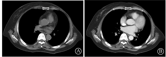
(1) CT定位和扫描:据报道[14],与胸式深吸气屏气相比,腹式深吸气屏气在降低危及器官受量方面更具有优势。因此,本研究纳入患者均采用腹式呼吸,患者定位前行2~3次呼吸训练,根据患者肺活量设定并记录吸气容积阈值,从而让患者适应美国医科达ABC系统装置。按照常规发泡胶+体膜固定、体位要求及体表标记后,配置ABC系统,使用荷兰飞利浦公司大孔径CT模拟机(Brilliance CT Big Bore)定位扫描,CT扫描范围从乳突平至肝脏的下缘,扫描层厚5 mm,所有患者采集2组CT定位图像,包括自由呼吸(free breath,FB)和DIBH(图 1)。

|
图 1 不同呼吸相上心脏相对位置的变化(第3肋间下缘) A. DIBH相图像;B. FB相图像 Figure 1 Relationship between chest wall and heart in different breathing modes (lower margin of the third intercostal) A. DIBH CT image; B. FB CT image |
(2) 靶区勾画与危及器官(organ at risk,OAR)勾画:在DIBH图像上勾画临床靶区体积(clinical target volume,CTV),范围包括左侧锁骨上下区、内乳区和胸壁,其中内乳区勾画至第3肋间(第4肋上缘),CTV外放0.5 cm生成计划靶区(planning target volume,PTV),且前界不超过皮肤表面下0.3 cm。在DIBH和FB两组CT图像上分别勾画包括心脏、双肺、肝、LAD、脊髓、甲状腺等。
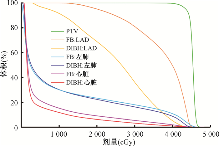
(3) 计划设计与优化:将CT定位图像上传至医科达公司Monaco(版本:5.11.03)放疗计划系统,在DIBH图像上制作放疗计划,计划设计采用6野动态调强技术(dynamic intensity-modulated radiotherapy,dIMRT)设计,能量为6 MV X射线,处方剂量是43.5 Gy/15次,单次剂量2.9 Gy。将FB和DIBH图像以治疗野内的乳房和胸壁进行匹配融合,根据融合的FB相上的OAR位置进行勾画并进行相应密度修改,据此来评估FB状态下OAR的剂量。比较并记录在DIBH及FB相状态下各患者全心脏、LAD及左肺的体积、平均剂量Dmean、最大剂量Dmax以及V5 ~ V30(Vx表示接收x Gy剂量照射的体积百分比),见图 2。

|
注:PTV. 计划靶区;FB. 自由呼吸;DIBH. 深吸气屏气;LAD. 冠状动脉左前降支 图 2 PTV和不同时相危及器官(左肺、心脏、LAD)的剂量体积直方图 Figure 2 Dose and volume histogram of PTV and organs at risk (left lung, heart, LAD) in different breathing modes |
(4) 治疗期间的图像验证:治疗前采用四维锥形束CT(Cone Beam CT,4D CBCT)扫描,用其最大吸气相与定位时DIBH时相的图像进行匹配和验证,验证频率为前3次放疗每次验证,之后每周2次。
4.统计学处理:对连续变量的数据结果以x±s表示,采用SPSS 26.0软件进行统计分析,采用K-S法对剂量体积参数行正态性检验,选择采用配对样本t检验和非参数Wilcoxion秩和检验,双变量相关分析采用Pearson检验,P<0.05为差异具有统计学意义。
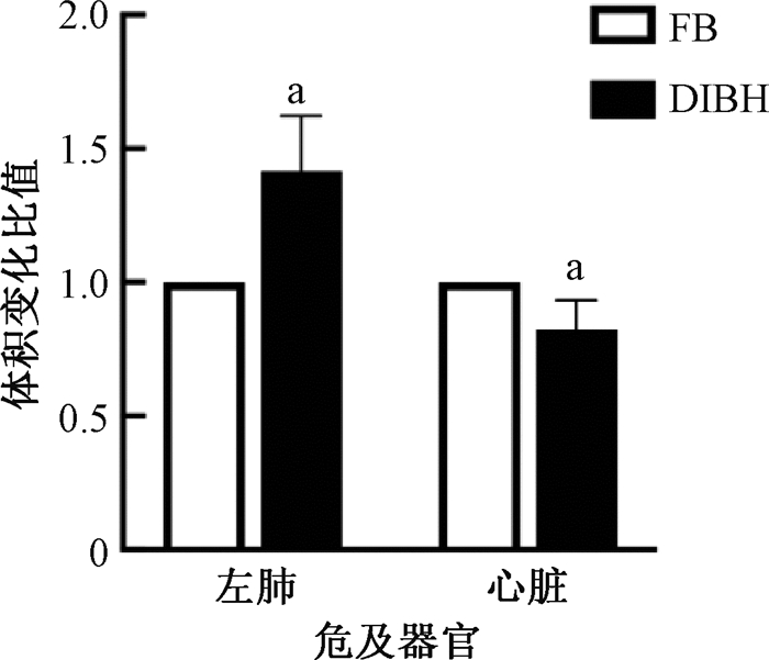
结果1.不同呼吸状态下心脏和肺的体积变化:与FB相比,DIBH状态下心脏体积缩小18% [(526.0±106.7)cm3 vs. (642.7±123.5)cm3,t= 10.47,P<0.001]、左肺体积扩张42%[(1 787.4 ± 286.6)cm3 vs. (1 223.7±211.0)cm3,t=-14.55,P<0.001,图 3]。

|
注:FB.自由呼吸;DIBH.深吸气屏气 a与FB组比较,t=14.55、-10.47,P<0.001 图 3 DIBH相上的患者左肺及心脏体积变化比值 Figure 3 Volume change ratios of left lung and heart in DIBH-treated patients |
2.不同呼吸状态下心、肺和LAD的剂量-体积参数的变化:与FB相比,DIBH状态下心脏及LAD平均剂量Dmean、最大剂量Dmax、剂量-体积参数V5 ~V30均明显降低(t=-13.38 ~ -3.30,P<0.05);心脏的Dmean减少(204.0±94.5)cGy,相对下降34%,剂量-体积参数V5、V10、V20、V30分别下降26.2%、35.0%、37.1%、63.1%;LAD的Dmean减少(1 123.2±375.4)cGy,相对下降37%;剂量-体积参数V5、V10、V20、V30分别下降8.2%、21.7%、45.9%、65.4%。左肺Dmean和V5也有所降低,分别相对下降3.76%和3.68%,差异具有统计学意义(t=-13.38~-2.60,P<0.05,表 2)。
|
|
表 2 心脏、冠状动脉左前降支及肺在DIBH及FB相上的剂量参数的比较(x±s) Table 2 Comparison of dosimetric parameters for the heart, left anterior descending coronary artery, and left lung between DIBH and FB modes (x±s) |
3.DIBH降低心脏剂量的影响因素分析:Pearson相关性分析提示,左肺扩张的相对比值与心脏剂量减少的相对比值(DFB-DDIBH)/ DFB呈正相关(r=0.82,P<0.001),但与肺剂量减少的相对比值无明显关系(r=0.16,P=0.462)。患者年龄与相对心脏剂量减少的相对比值呈负相关(r=-0.57,P=0.005,图 4)。

|
图 4 深吸气屏气患者心脏剂量减少的相对比值与左肺扩张的相对比值(A)及患者年龄(B)的相关性 Figure 4 Correlation between the relative ratio of cardiar dose reduction and the relative ratio of left lung expansion(A) and patient age(B) |
讨论
放疗时心脏的辐射暴露是其损伤的主要因素,由于心脏位于左侧胸腔,一般情况下,左侧乳腺癌术后放疗的患者,其心脏受量明显高于右侧乳腺癌术后放疗者,并且其心脏病的发生风险也随之增加[15]。陈偲晔等[16]研究表明,左侧乳腺癌保乳术后采用DIBH行全乳IMRT可以有效地降低心脏剂量。本研究也证实在照射范围更大的IMNI中实施DIBH,可以明显降低心脏及其亚结构的剂量。因此,IMNI患者也可推荐行DIBH技术进行心脏保护。
IMRT技术作为精确放疗的代表,已在临床广泛应用。一项对比左侧乳腺癌术后IMRT和三维适形放疗(three dimensional radiation therapy,3D-CRT)剂量学的研究提示[17],IMRT计划靶区的适形度(conformity index,CI)和剂量的均匀性(homogeneity index,HI),以及在患侧肺、心脏和左心室的高剂量体积参数均优于3D-CRT计划,但上述危及器官的低剂量体积参数则劣于3D-CRT计划。由于引起心脏损伤的辐照剂量并没有明显的阈值,即使是低剂量区也可能对心脏产生损伤[18],因此,如何降低心脏及亚结构低剂量区也值得关注。本研究发现,在IMRT的基础上实施DIBH,可以有效的降低心脏及其亚结构LAD的V5、V10等低剂量体积参数。此外,DIBH技术本身延长了患者每次治疗的时间,给放疗计划实施的精准度带来了潜在的干扰。随着放疗设备和软件的更新和发展,容积弧形调强放疗(volumetric modulated arc therapy,VMAT)技术凭借其精准、高效的特点得到了广泛的推广和应用,其相较于IMRT技术可以有效缩短放疗时间[19],但是其对左乳癌患者心脏剂量的影响,结论尚存争议[20-21],还需要更进一步探索和研究。
由于LAD的解剖学特点,其走行范围贴近左侧胸壁,因此左侧乳腺癌术后放疗时,LAD受照剂量高,并且其损伤以及狭窄程度与所受辐照剂量密切相关[22-24]。多项研究表明,DIBH技术可以有效降低左侧乳腺癌术后放疗时的心脏受量,相对于FB时相而言,DIBH时心脏平均剂量可减少40%~50%,LAD平均剂量降低43.5% ~72.1%[25-27]。本研究中,DIBH时相心脏及LAD剂量的降低幅度均小于以往的数据,LAD平均剂量的最大降幅也仅为37%。这可能与纳入人群的均为IMNI有关,更大范围的内乳靶区造成了更高的心脏剂量,从而削弱了DIBH技术在心脏剂量学上的改善作用。
关于DIBH对肺部受量的影响,既往研究结果并不一致,有研究发现DIBH技术可以使同侧肺V20降低约10% ~ 30%[28],而另一些研究提示DIBH和FB状态下并没有明显差别甚至会升高[26, 29]。本研究则发现,DIBH能够减少同侧肺的Dmean和V5,而对其V20及V30无明显降低,这可能与包含内乳的照射范围其切线野内有更多的肺组织,从而造成深吸气肺部扩张时高剂量区减少不明显有关。因此,DIBH对左乳癌术后IMNI-IMRT肺部受量的影响尚需利用更多的临床数据加以明确。此外,本研究与其他报道一致[28],进一步确认了肺扩张的程度与心脏剂量减少的程度显著相关,从而提示在DIBH实施过程中,尽力吸气屏气会更有助于心脏剂量的降低。本研究还发现年龄与心脏剂量减少程度密切相关,即年龄越轻,心脏剂量减少程度越大。这可能与年轻女性患者肺活量大、吸气时纵隔及心脏运动幅度相对较大有关。
本研究进一步明确了DIBH技术联合IMRT在降低左侧乳腺癌IMNI患者心脏及其相关亚结构剂量上的优势,而该技术是否能真正减少放疗后心脏损伤的发生,还需要长期的临床随访来验证。
利益冲突 无
作者贡献声明 周咏春负责研究设计和论文撰写;孙祥露、吴焕参与论文撰写;孙楠负责数据收集;李威负责数据分析;韩洋负责技术流程的实施;邓虎、谢凌霄、张蕾、符诗薇协助数据分析;江浩指导研究设计
| [1] |
Xia C, Dong X, Li H, et al. Cancer statistics in china and united states, 2022: Profiles, trends, and determinants[J]. Chin Med J (Engl), 2022, 135(5): 584-590. DOI:10.1097/CM9.0000000000002108 |
| [2] |
Abdel-Qadir H, Austin PC, Lee DS, et al. A population-based study of cardiovascular mortality following early-stage breast cancer[J]. JAMA Cardiol, 2017, 2(1): 88-93. DOI:10.1001/jamacardio.2016.3841 |
| [3] |
Darby SC, Ewertz M, McGale P, et al. Risk of ischemic heart disease in women after radiotherapy for breast cancer[J]. N Engl J Med, 2013, 368(11): 987-998. DOI:10.1056/NEJMoa1209825 |
| [4] |
Clarke M, Collins R, Darby S, et al. Effects of radiotherapy and of differences in the extent of surgery for early breast cancer on local recurrence and 15-year survival: an overview of the randomised trials[J]. Lancet, 2005, 366(9503): 2087-2106. DOI:10.1016/S0140-6736(05)67887-7 |
| [5] |
Laugaard Lorenzen E, Christian Rehammar J, Jensen MB, et al. Radiation-induced risk of ischemic heart disease following breast cancer radiotherapy in Denmark, 1977-2005[J]. Radiother Oncol, 2020, 152: 103-110. DOI:10.1016/j.radonc.2020.08.007 |
| [6] |
Henson KE, McGale P, Taylor C, et al. Radiation-related mortality from heart disease and lung cancer more than 20 years after radiotherapy for breast cancer[J]. Br J Cancer, 2013, 108(1): 179-182. DOI:10.1038/bjc.2012.575 |
| [7] |
Taylor C, Correa C, Duane FK, et al. Estimating the risks of breast cancer radiotherapy: Evidence from modern radiation doses to the lungs and heart and from previous randomized trials[J]. J Clin Oncol, 2017, 35(15): 1641-1649. DOI:10.1200/JCO.2016.72.0722 |
| [8] |
van den Bogaard VA, Ta BD, van der Schaaf A, et al. Validation and modification of a prediction model for acute cardiac events in patients with breast cancer treated with radiotherapy based on three-dimensional dose distributions to cardiac substructures[J]. J Clin Oncol, 2017, 35(11): 1171-1178. DOI:10.1200/JCO.2016.69.8480 |
| [9] |
Duma MN, Baumann R, Budach W, et al. Heart-sparing radiotherapy techniques in breast cancer patients: a recommendation of the breast cancer expert panel of the German society of radiation oncology (DEGRO)[J]. Strahlenther Onkol, 2019, 195(10): 861-871. DOI:10.1007/s00066-019-01495-w |
| [10] |
Lai J, Hu S, Luo Y, et al. Meta-analysis of deep inspiration breath hold (DIBH) versus free breathing (FB) in postoperative radiotherapy for left-side breast cancer[J]. Breast Cancer, 2020, 27(2): 299-307. DOI:10.1007/s12282-019-01023-9 |
| [11] |
Kim YB, Byun HK, Kim DY, et al. Effect of elective internal mammary node irradiation on disease-free survival in women with node-positive breast cancer: A randomized phase 3 clinical trial[J]. JAMA Oncol, 2022, 8(1): 96-105. DOI:10.1001/jamaoncol.2021.6036 |
| [12] |
Thorsen L, Overgaard J, Matthiessen LW, et al. Internal mammary node irradiation in patients with node-positive early breast cancer: Fifteen-year results from the danish breast cancer group internal mammary node study[J]. J Clin Oncol, 2022, 40(36): 4198-4206. DOI:10.1200/JCO.22.00044 |
| [13] |
Taylor CW, Wang Z, Macaulay E, et al. Exposure of the heart in breast cancer radiation therapy: A systematic review of heart doses published during 2003 to 2013[J]. Int J Radiat Oncol Biol Phys, 2015, 93(4): 845-853. DOI:10.1016/j.ijrobp.2015.07.2292 |
| [14] |
Zhao F, Shen J, Lu Z, et al. Abdominal DIBH reduces the cardiac dose even further: a prospective analysis[J]. Radiat Oncol, 2018, 13(1): 116. DOI:10.1186/s13014-018-1062-6 |
| [15] |
Rehammar JC, Jensen MB, McGale P, et al. Risk of heart disease in relation to radiotherapy and chemotherapy with anthracyclines among 19, 464 breast cancer patients in Denmark, 1977-2005[J]. Radiother Oncol, 2017, 123(2): 299-305. DOI:10.1016/j.radonc.2017.03.012 |
| [16] |
陈偲晔, 王淑莲, 唐玉, 等. 左侧乳腺癌保乳术后采用深吸气屏气放疗的心脏剂量学分析[J]. 中华放射肿瘤学杂志, 2018, 27(3): 281-288. Chen SY, Wang SL, Tang Y, et al. Cardiac dosimetriy of deep inspiration breath hold technique in whole breast irradiation for left breast cancer after breast irradiation for left breast cancer after breast conserving surgery[J]. Chin J Radiat Oncol, 2018, 27(3): 281-288. DOI:10.3760/cma.j.issn.1004-4221.2018.03.011 |
| [17] |
魏世鸿, 陶娜, 欧阳水根, 等. 左侧乳腺癌根治术后IMRT和3D-CRT放疗技术剂量学比较[J]. 中华肿瘤防治杂志, 2019, 26(12): 855-860. Wei SH, Tao N, Ouyang SG, et al. Dosimetric comparison of IMRT and 3D-CRT radiotherapy for left-sided breast cancer after radical surgery[J]. Chin J Cancer Prev Treat, 2019, 26(12): 855-860. DOI:10.16073/j.cnki.cjcpt.2019.12.007 |
| [18] |
Milder CM, Kendall GM, Arsham A, et al. Summary of Radiation Research Society Online 66th Annual Meeting, Symposium on "Epidemiology: Updates on epidemiological low dose studies, " including discussion[J]. Int J Radiat Biol, 2021, 97(6): 866-873. DOI:10.1080/09553002.2020.1867326 |
| [19] |
Liu H, Chen X, He Z, et al. Evaluation of 3D-CRT, IMRT and VMAT radiotherapy plans for left breast cancer based on clinical dosimetric study[J]. Comput Med Imaging Graph, 2016, 54: 1-5. DOI:10.1016/j.compmedimag.2016.10.001 |
| [20] |
Nair SS, Devi V, Sharan K, et al. A dosimetric study comparing different radiotherapy planning techniques with and without deep inspiratory breath hold for breast cancer[J]. Cancer Manag Res, 2022, 14: 3581-3587. DOI:10.2147/CMAR.S381316 |
| [21] |
Sakka M, Kunzelmann L, Metzger M, et al. Cardiac dose-sparing effects of deep-inspiration breath-hold in left breast irradiation: Is IMRT more beneficial than VMAT?[J]. Strahlenther Onkol, 2017, 193(10): 800-811. DOI:10.1007/s00066-017-1167-0 |
| [22] |
Taylor C, McGale P, Brønnum D, et al. Cardiac structure injury after radiotherapy for breast cancer: Cross-sectional study with individual patient data[J]. J Clin Oncol, 2018, 36(22): 2288-2296. DOI:10.1200/JCO.2017.77.6351 |
| [23] |
Nilsson G, Holmberg L, Garmo H, et al. Distribution of coronary artery stenosis after radiation for breast cancer[J]. J Clin Oncol, 2012, 30(4): 380-386. DOI:10.1200/JCO.2011.34.5900 |
| [24] |
Wennstig AK, Garmo H, Isacsson U, et al. The relationship between radiation doses to coronary arteries and location of coronary stenosis requiring intervention in breast cancer survivors[J]. Radiat Oncol, 2019, 14(1): 40. DOI:10.1186/s13014-019-1242-z |
| [25] |
Menezes KM, Wang H, Hada M, et al. Radiation matters of the heart: A mini review[J]. Front Cardiovasc Med, 2018, 5: 83. DOI:10.3389/fcvm.2018.00083 |
| [26] |
Yeung R, Conroy L, Long K, et al. Cardiac dose reduction with deep inspiration breath hold for left-sided breast cancer radiotherapy patients with and without regional nodal irradiation[J]. Radiat Oncol, 2015, 10: 200. DOI:10.1186/s13014-015-0511-8 |
| [27] |
Salvestrini V, Iorio GC, Borghetti P, et al. The impact of modern radiotherapy on long-term cardiac sequelae in breast cancer survivor: a focus on deep inspiration breath-hold (DIBH) technique[J]. J Cancer Res Clin Oncol, 2022, 148(2): 409-417. DOI:10.1007/s00432-021-03875-1 |
| [28] |
Song J, Tang T, Caudrelier JM, et al. Dose-sparing effect of deep inspiration breath hold technique on coronary artery and left ventricle segments in treatment of breast cancer[J]. Radiother Oncol, 2021, 154: 101-109. DOI:10.1016/j.radonc.2020.09.019 |
| [29] |
Rice L, Goldsmith C, Green MM, et al. An effective deep-inspiration breath-hold radiotherapy technique for left-breast cancer: impact of post-mastectomy treatment, nodal coverage, and dose schedule on organs at risk[J]. Breast Cancer (Dove Med Press), 2017, 9: 437-446. DOI:10.2147/BCTT.S130090 |
 2023, Vol. 43


