2. 瓦里安医疗设备(中国)有限公司, 北京 100176
2. Varian Medical System, Beijing 100176, China
近些年来,图像引导的放射治疗(imageguided radiation therapy,IGRT)的快速发展进一步提高了患者摆位和靶区定位的精度[1-4]。锥形束CT(cone-beam CT,CBCT)在监测内部器官运动、形变及外轮廓改变等方面具有独特的优势[5-6]。但是与常规扇形束CT图像相比,无论空间分辨率还是密度分辨率都有一定的差距[7-9]。迭代锥形束CT(iterative cone-beam CT,ICBCT)是近年来在CBCT图像基础上,通过迭代重建图像噪音降低算法发展起来的一项新技术,与常规CBCT图像相比,图像质量明显提高[10-13]。本文的目的是研究利用盆腔迭代CBCT图像用于治疗计划剂量计算的可行性分析,为自适应放疗提供图像保障。
资料与方法 1、锥形束CT-电子密度转换曲线(iterative Cone-beam CT to electron density,ICBCT-ED)的建立在Varian Halcyon 2.0环形加速器上,利用美国CIRS公司的062 M专用模体(CIRS Tissue Simulation & Phantom Technology, Norfolk, VA, USA)建立不同散射条件下ICBCT-ED转换曲线。为验证不同散射条件下CT值的差异,共模拟14种模体摆放位置。分别为11种头脚方向各增加30 cm×30 cm×30 cm固体水的全散射位置(中心摆放、左右移动5 cm、左右移动10 cm、前后移动5 cm、前后移动10 cm、仅包含内圈、仅包含外圈);两种头脚方向包含5和10 cm固体水的半散射位置;1种无散射体的孤立模式。Halcyon kvCBCT图像采集采用“pelvis”模式(125 kV、30 mA、10 ms),重建层厚2 mm,重建长度24.5 cm。求其不同散射条件下的CT平均值,在Eclipse 15.6计划系统中建立与之对应的ICBCT-ED转换曲线。
2、基于CIRS盆腔模体CT图像和ICBCT图像的剂量学比较使用Phillips Brilliance CT (荷兰,飞利浦公司)采集美国CIRS公司的002PRA盆腔调强专用模体(CIRS Tissue 10Simulation & Phantom Technology, Norfolk, VA, USA)的CT图像,扫描条件(140 kV、325 mAs), 层厚2 mm。传输图像至Eclipse计划系统,在盆腔模体CT图像上按照宫颈癌术后患者勾画CTV、PTV、膀胱、直肠、小肠、骨髓、股骨头、脊髓等危及器官,设计4弧VMAT计划,处方剂量45 Gy/25次,射线能量6 MV,剂量率800 MU/min,剂量计算采用AAA模型。
在Halcyon加速器,按照“pelvis”模式,采集盆腔模体位于4种位置处(左右移动5 cm、左右移动10 cm)的ICBCT图像,拷贝基于CT图像上的靶区和危及器官并移植与之对应的4弧VMAT计划至ICBCT图像上,应用ICBCT-ED转换曲线,重新进行剂量计算。利用剂量体积直方图评估靶区及危及器官的剂量差异。使用PTW verisoft 7.1版本软件(PTW,Frieburg, Germany)分析三维体积剂量的γ通过率,评估标准1%/1 mm、2%/2 mm、3%/3 mm,阈值10%。
3、临床患者CT图像和ICBCT图像的剂量学回顾性分析随机抽样2020年12月北京协和医院治疗的10例盆腔肿瘤患者,其中宫颈癌术后患者3例(28次)、宫颈癌根治术后3例(20次)、直肠癌新辅助患者4例(25次),所有患者每次均行ICBCT图像采集。拷贝靶区和危及器官至每次的ICBCT图像,重新勾画外轮廓,移植与之对应的患者治疗计划并重新进行剂量再计算,使用PTW verisoft软件分析每次三维体积剂量的γ通过率,评估标准2%/2 mm、3%/3 mm,阈值10%。
结果 1、不同散射条件下ICBCT-ED转换曲线的差异表 1为不同散射条件CT值与ED的对应关系。由数据分析可知,两种全散射位置(仅包含内圈、仅包含外圈);两种半散射位置;1种无散射体的孤立模式与全散射中心位置的CT值偏差较大,最大偏差144 HU。其他全散射位置与中心位置CT值相近,最大偏差 < 50 HU。故在Eclipse计划系统拟合ICBCT-ED转换曲线需采用离散性较小的9条曲线的平均值,拟合曲线与中心曲线相比,最大偏差 < 10 HU。
|
|
表 1 不同散射条件下不同电子密度对应值(HU-g/cm3) Table 1 ICBCT-ED value under different scattering conditions(HU-g/cm3) |
2、CIRS盆腔模体不同扫描位置处靶区、危及器官及三维剂量γ通过率的差异
基于盆腔模体不同位置处的ICBCT图像的的计算结果,与基于CT图像的计划相比,无论靶区还是危及器官的剂量偏差均 < 1 Gy(表 2,3)。与基于CT图像的计划相比,基于ICBCT图像的三维剂量γ通过率如表 4所示,1%/1 mm和2%/2 mm的平均值分别为(88.86±1.18)%和(98.38±0.89)%。
|
|
表 2 盆腔模体不同位置处靶区剂量参数(Gy) Table 2 Dose parameters for target area at different positions of pelvic phantom (Gy) |
|
|
表 3 盆腔模体不同位置处危及器官剂量参数(Gy) Table 3 Dose parameters for organs at risk at different positions of pelvic phantom (Gy) |
|
|
表 4 盆腔模体不同位置处三维剂量γ通过率(%) Table 4 Three dimensional pantom-based gamma dose passing rate at different positions of pelvic phantom (%) |
3、盆腔患者不同分次间三维剂量γ通过率的差异
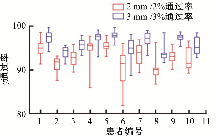
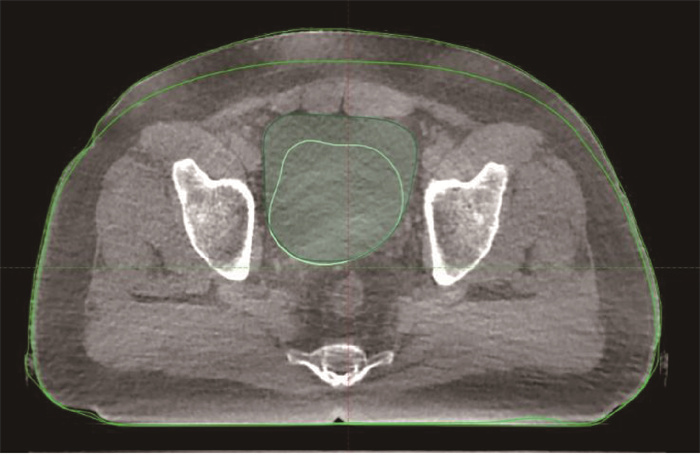
10例盆腔肿瘤患者基于ICBCT图像的三维剂量通过率如图 1所示。10例盆腔肿瘤患者2%/2 mm和3%/3 mm的平均值范围分别为90.03%~95.43%和93.58%~97.78%。最差结果如图 2所示,为患者6第17次治疗时由于膀胱过充盈引起的外轮廓变化,2%/2 mm和3%/3 mm的三维剂量通过率仅为85.90%和92.90%。

|
图 1 10例盆腔肿瘤患者的三维剂量γ通过率 Figure 1 Three-dimensional gamma dose passing rates of 10 patients |

|
图 2 盆腔肿瘤患者膀胱充盈状态对外轮廓的影响 Figure 2 Effect of bladder filling on external contour |
讨论
Halcyon加速器2.0版本的ICBCT盆腔采集模式有两种,为Pelvis和Pelvis Large,其头脚方向的理论重建长度分别为24.5和28 cm,鉴于图像重建完整性的原因,其实际可用于剂量再计算的长度仅为20和24 cm。这样就限制了ICBCT在自适应放疗中的应用范围。能否像Truebeam加速器一样开展扩展ICBCT功能建议厂家进一步研究,为后续的自适应放疗提供图像保障。
基于ICBCT的自适应放疗图像质量是关键,空腔散射伪影和运动伪影是影像图像质量的两个主要因素[14]。尤其前者,可导致CT值的偏差值达到100 HU以上,故如何降低空腔散射伪影对剂量计算精度的影响是下步研究的关键,这点从ICBCT-ED的转换曲线上也可以反应出来。Pelvis模式下,ICBCT的采集时间需要36.7 s,小肠等运动器官在图像采集周期内产生运动伪影也势必影响图像质量,但是与散射伪影相比,相对较小。
研究结果表明,当模体远离等中心位置时,CT值受位置的影响还是较大的,最大偏差达170 HU。但是相对电子密度偏差最大仅差0.1 g/cm3, 这对计算影响是非常有限的,遗憾是本研究并未对不同模体位置的采集图像进行剂量再计算和伽玛通过率比较[15]。但是,本实验结果表明当模体在左右方向有位置偏移时,CT值和三维剂量伽玛通过率均有着较好的一致性,主要原因可能与ICBCT的算法改进有一定的关系。从图 1的结果也可以看出,位置对ICBCT的CT值的影响较小,主要的影响来自于散射线的贡献。
本研究在利用ICBCT进行计划剂量再计算时有两个方面的不足,一是仅对盆腔部位的可行性分析进行了研究,而对其他部位还没有涉及;二是在实际患者剂量比较时只分析了三维剂量的γ通过率,而对危及器官的DVH没有评估。为此将进行基于头颈部ICBCT的剂量学可行性分析,以及基于Velocity不同分次的的剂量叠加等。
影响基于不同图像的三维剂量的差异的相关因素是多方面的。如体重变化引起的外轮廓的改变、器官充盈及伪影引起的电子密度差异、图像配准引起的等中心位置的变化均会导致三维剂量γ通过率的降低,故在利用ICBCT图像制定自适应放疗方案时要重点考虑这几个方面带来的影响[16-20]。
综上所述,与CBCT图像相比,ICBCT图像有了进一步的提高,在某些方面的图像质量接近扇形束CT。在足够的散射条件下,重建ICBCT-ED转换曲线,利用Halcyon直线加速器的ICBCT图像进行治疗计划设计,其精度是可以满足临床应用的标准的,为将来的自适应放疗提供了保障。
利益冲突 所有作者声明不存在利益冲突
作者贡献声明 杨波负责设计研究方案和论文撰写;汪之群、祝起禛参与测量、收集和整理数据;李文博、黎蕊协助数据统计分析;张新、潘俊生提供技术支持;胡克、张福泉和邱杰提供科研思路和科研方案指导
| [1] |
Li T, Schreibmann E, Yang Y, et al. Motion correction for improved target localization with on-board cone-beam computed tomography[J]. Phys Med Biol, 2006, 51(2): 253-267. DOI:10.1088/0031-9155/51/2/005 |
| [2] |
Purdie TG, Bissonnette JP, Franks K, et al. Cone-beam computed tomography for on-line image guidance of lung stereotactic radiotherapy: localization, verification, and intrafraction tumor position[J]. Int J Radiat Oncol Biol Phys, 2007, 68(1): 243-252. DOI:10.1016/j.ijrobp.2006.12.022 |
| [3] |
Franzone P, Fiorentino A, Barra S, et al. Image-guided radiation therapy (IGRT): practical recommendations of Italian Association of Radiation Oncology (AIRO)[J]. Radiol Med, 2016, 121(12): 958-965. DOI:10.1007/s11547-016-0674-x |
| [4] |
Alongi F, de Crevoisier R, Corradini S, et al. Daily IGRT for prostate cancer: Can we stop the train?[J]. Radiother Oncol, 2018, 128(2): 389-390. DOI:10.1016/j.radonc.2018.05.009 |
| [5] |
Létourneau D, Wong JW, Oldham M, et al. Cone-beam-CT guided radiation therapy: technical implementation[J]. Radiother Oncol, 2005, 75(3): 279-286. DOI:10.1016/j.radonc.2005.03.001 |
| [6] |
Nakagawa K, Yamashita H, Shiraishi K, et al. Verification of in-treatment tumor position using kilovoltage cone-beam computed tomography: a preliminary study[J]. Int J Radiat Oncol Biol Phys, 2007, 69(4): 970-973. DOI:10.1016/j.ijrobp.2007.08.026 |
| [7] |
Sorcini B, Tilikidis A. Clinical application of image-guided radiotherapy, IGRT (on the Varian OBI platform)[J]. Cancer Radiother, 2006, 10(5): 252-257. DOI:10.1016/j.canrad.2006.05.012 |
| [8] |
Taneja S, Barbee DL, Rea AJ, et al. CBCT image quality QA: Establishing a quantitative program[J]. J Appl Clin Med Phys, 2020, 21(11): 215-225. DOI:10.1002/acm2.13062 |
| [9] |
杨波, 邱杰, 庞廷田, 等. 头颈部CBCT图像对放疗剂量计算准确性的影响[J]. 中华放射医学与防护杂志, 2008, 28(5): 523-525. Yang B, Qiu J, Pang TT, et al. Influence of CBCT image of head and neck on accuracy of radiotherapy dose calculation[J]. Chin J Radiol Med Prot, 2008, 28(5): 523-525. DOI:10.3760/cma.j.issn.0254-5098.2008.05.025 |
| [10] |
Cai B, Laugeman E, Mazur TR, et al. Characterization of a prototype rapid kilovoltage X-ray image guidance system designed for a ring shape radiation therapy unit[J]. Med Phys, 2019, 46(3): 1355-1370. DOI:10.1002/mp.13396 |
| [11] |
Gardner SJ, Mao W, Liu C, et al. Improvements in CBCT image quality using a novel iterative reconstruction algorithm: a clinical evaluation[J]. Adv Radiat Oncol, 2019, 4(2): 390-400. DOI:10.1016/j.adro.2018.12.003 |
| [12] |
Hashemi S, Song WY, Sahgal A, et al. Simultaneous deblurring and iterative reconstruction of CBCT for image guided brain radiosurgery[J]. Phys Med Biol, 2017, 62(7): 2521-2541. DOI:10.1088/1361-6560/aa5ed2 |
| [13] |
Washio H, Ohira S, Funama Y, et al. Metal artifact reduction using iterative CBCT reconstruction algorithm for head and neck radiation therapy: a phantom and clinical study[J]. Eur J Radiol, 2020, 132: 109293. DOI:10.1016/j.ejrad.2020.109293 |
| [14] |
Zhu L, Xie Y, Wang J, et al. Scatter correction for cone-beam CT in radiation therapy[J]. Med Phys, 2009, 36(6): 2258-2268. DOI:10.1118/1.3130047 |
| [15] |
Jarema T, Aland T. Using the iterative kV CBCT reconstruction on the Varian Halcyon linear accelerator for radiation therapy planning for pelvis patients[J]. Phys Med, 2019, 68: 112-116. DOI:10.1016/j.ejmp.2019.11.015 |
| [16] |
Barsky AR, Kim MM, Dreyfuss A, et al. Initial clinical experience treating patients with gynecologic cancers on a 6 MV flattening filter free o-ring linear accelerator[J]. Adv Radiat Oncol, 2020, 5(5): 920-928. DOI:10.1016/j.adro.2020.01.004 |
| [17] |
Sonke JJ, Aznar M, Rasch C. Adaptive radiotherapy for anatomical changes[J]. Semin Radiat Oncol, 2019, 29(3): 245-257. DOI:10.1016/j.semradonc.2019.02.007 |
| [18] |
Frcr L, Tanderup K, Kirisits C, et al. Image-guided adaptive radiotherapy in cervical cancer[J]. Semin Radiat Oncol, 2019, 29(3): 284-298. DOI:10.1016/j.semradonc.2019.02.010 |
| [19] |
Westerveld H, Nesvacil N, Fokdal L, et al. Definitive radiotherapy with image-guided adaptive brachytherapy for primary vaginal cancer[J]. Lancet Oncol, 2020, 21(3): e157-157e167. DOI:10.1016/S1470-2045(19)30855-1 |
| [20] |
Briens A, Castelli J, Barateau A, et al. Adaptive radiotherapy: Strategies and benefits depending on tumor localization[J]. Cancer Radiother, 2019, 23(6-7): 592-608. DOI:10.1016/j.canrad.2019.07.135 |
 2021, Vol. 41


