2. 复旦大学核物理和离子束应用教育部重点实验室, 上海 200433
2. Key Laboratory of Nuclear Physics and Ion-beam Application(MOE), Fudan University, Shanghai 200433, China
固体核径迹探测材料聚丙烯二甘醇碳酸脂(C12H18O7),俗称CR-39,作为被动式的电离辐射探测器,时常被推荐用来测量带电粒子,如质子、α粒子、重离子等[1-3]。其原理是带电粒子入射到CR-39时,在沿着粒子路径方向上的能量沉积会造成CR-39材料的损伤,形成潜径迹[4],而这样的潜径迹经化学或电化学蚀刻后,会生长成微米级径迹,可由光学显微镜、扫描电镜或原子力显微镜等[5-7]装置观测到。这种可视的信号对电离辐射测量,无疑具有重要的研究和应用价值。
近十余年来,随着核技术及其应用的不断蓬勃发展,越来越多的领域需要开展质子的测量及其辐射防护与剂量学研究,如质子和中子放射治疗、激光驱动质子加速器研发、核聚变研究等[8-10]。因此,关于CR-39对质子的主要探测特性-能量与角度响应,已有不少研究者开展了相关研究[11-13]。但是,到目前为止,绝大部分研究都是在探究CR-39可响应能量范围和可探测到的最大入射角度(临界角)的问题,鲜见有比较详尽的、利用核径迹来估算入射质子的能量和角度的研究。本研究通过用不同入射能量和入射角度的质子开展实验,并借助于固体核径迹二维形态,旨在探索建立基于核径迹形态来估算入射质子的能量和角度的方法。
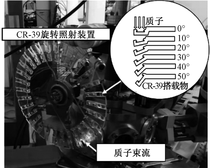
材料与方法1.照射实验:利用复旦大学现代物理研究所的9SDH-2串列加速器(美国NEC公司产品)[14],在最小束流条件下产生能量在1.9~3.3 MeV范围内的5种单能质子,将5组共30片CR-39(Fukuvi Chemical Industry Co., Ltd)放在一个自制的可旋转支架上尽量贴近射束出口进行照射。为提高实验效率,旋转支架上放置CR-39的凹槽被设计为不同形状,以模拟入射角度为0°~50°的照射条件。为确保入射质子在CR-39上形成密度适中的核径迹(不太少、也尽量不发生重叠),支架的旋转速度由多次预实验结果来设定。照射实验的现场照片如图 1所示。为更加准确定量入射到CR-39表面的质子能量,考虑到射束出口膜的阻挡和空气的衰减,借助GEANT 4®蒙特卡罗模拟软件,分别计算了不同照射条件下质子入射到CR-39表面上的能量和质子在CR-39中的射程,结果如表 1所示。

|
图 1 质子照射实验照片 Figure 1 A photo of proton irradiation experiment |
|
|
表 1 入射到CR-39上的质子能量和射程 Table 1 Incident energies of protons and their ranges in CR-39 |
2.CR-39蚀刻和径迹形态读取:基于预实验结果,被质子照射后的CR-39统一用6.25 N的氢氧化钠溶液,在(90±0.1)℃条件下进行4 h化学蚀刻。在上述蚀刻条件下,通过重量法[15]测得体蚀刻速率Vb为(5.61±0.11)μm/h。为分析不同照射条件下生成核径迹的二维形态,采用自主研发的核径迹自动读取装置(分辨率为0.32 μm/pixel)对核径迹的长、短轴进行自动定量分析。为进一步验证自主研发装置分析结果的可靠性,本研究还借助了上海交通大学分析测试中心的原子力显微镜(AFM,分辨率为0.08 μm/pixel)对部分核径迹的长、短轴分析结果进行验证。
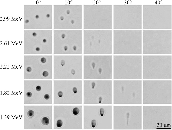
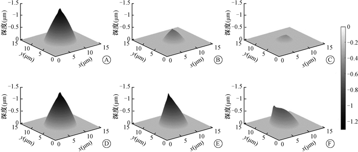
结果1.径迹形态学特性:在本实验研究的化学蚀刻条件下,图 2给出了在光学显微镜(×40)上可看到不同入射能量和入射角度的质子在CR-39上形成核径迹的典型图像。从图 2可以看出,随着入射能量的增加,核径迹的大小和光学对比度逐渐减小;不同入射角度质子形成的核径迹开口形状不同,垂直入射的质子的核径迹开口多呈圆形,而其他角度入射质子的核径迹开口多呈椭圆形状。对于1.39 MeV的入射质子,当入射角度>40°时,无法从显微镜中识别核径迹,随着入射质子能量的升高,可观测到的不同角度入射粒子的信息越来越少。图 3给出了利用AFM测量得到不同入射能量和入射角度的质子在CR-39上形成核径迹的典型图像。从图 3可以看出,所有径迹均呈仿锥形形状;高能质子形成的核径迹相对较小、深度也较浅。这可以解释为高能质子的传能线密度(LET)值较小,在穿过路径上损失的能量较小,从而径迹生长较慢。

|
图 2 不同入射能量和入射角度质子形成径迹的图像 Figure 2 Track images produced by different energies and angles of incident protons |

|
图 3 质子径迹三维图像 A. 1.39 MeV, 0°;B. 2.22 MeV, 0°;C. 2.99 MeV, 0°;D. 1.39 MeV, 0°;E. 1.39 MeV, 10°;F. 1.39 MeV, 20° Figure 3 3D images of proton tracks A. 1.39 MeV, 0°; B. 2.22 MeV, 0°; C. 2.99 MeV, 0°; D. 1.39 MeV, 0°; E. 1.39 MeV, 10°; F. 1.39 MeV, 20° |
2.两种读取方法获得核径迹的形态比较:在不同入射能量和角度的质子照射条件下,本研究同时用光学显微镜和AFM分析了它们在CR-39上形成核径迹的形态。利用简单随机抽样方法选择每种照射条件下的CR-39各1片,表 2汇总了用光学显微镜随机分析其中500个径迹和用AFM随机分析其中50个径迹的形态学信息。从表 2可以看出,总体来说,光学显微镜自动分析得到核径迹的长、短轴和用AFM测量得到的长、短轴基本相同,但也发现光学显微镜的分析结果平均比AFM的测值偏大约5.5%(1.9%~9.5%),核径迹越深、偏大越多。这可能是因为光学显微镜的散射光和光学系统的对焦偏移导致了核径迹边缘出现伪影,越深的径迹对散射和光学系统对焦会有更大的影响。
|
|
表 2 光学显微镜和原子力显微镜测量核径迹的形态比较(x±s) Table 2 Morphological information of tracks by optical microscope and AFM(x±s) |
3.质子能量与核径迹尺寸关系:考虑到用光学显微镜分析的径迹更多,图 4展示了在垂直入射和10°入射条件下,不同入射能量的质子与其形成核径迹的长、短轴以及等效直径的分布情况。借助于Origin软件采用基于Levernberg-Marquardt算法(LMA)的非线性最小二乘法拟合,发现入射质子的能量与核径迹的长、短轴或等效直径均符合很好的指数衰减关系(E=Ae-Bx),决定系数(R2)与核径迹短轴长度的关系最接近于1。上述结果反映了可通过测量核径迹的短轴来估算入射质子的能量。

|
图 4 径迹尺寸与能量分布及其拟合结果 A.短轴;B.长轴;C.等效直径 Figure 4 Distribution of track size and energy of incident proton and their fitting results A. Minor axis; B. Major axis; C. Equivalent diameter |
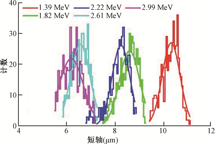
图 5为在入射角度为0°时不同入射能量质子在CR-39上形成核径迹的短轴长度分布。经高斯拟合,得到各分布图的半高宽分别为1.190 6、0.954 8、0.870 8、0.999 5和0.882 1 μm。经计算,对1.39、1.82、2.22、2.61和2.99 MeV入射质子能量的分辨率分别可达8.5%、11.6%、10.5%、14.5%和19.2%。

|
图 5 不同入射能量质子形成径迹的短轴分布 Figure 5 Distribution of minor axis of tracks produced by protons with different energies |
4.质子入射角度与核径迹长短轴比的关系:由表 2可知,随着入射质子角度的增大,核径迹的长短轴之比也随之增大。这说明了可望通过测量核径迹的长短轴之比来估算入射质子的角度。对垂直入射情况,核径迹长短轴之比的均值为1.03~1.08,反映了核径迹开口均接近圆形;对10°入射的质子,核径迹长短轴之比的均值为1.10~1.15;对20°入射的质子,核径迹长短轴之比的均值为1.35~1.37。上述结果表明,在本实验研究条件下,若利用核径迹的长短轴之比已可区分差异在10°左右的入射质子。
讨论CR-39在质子测量中的应用越来越广泛,深入研究其对入射质子的能量和角度的分辨能力,无疑对各种辐射场中质子的能谱解析、准确计算其在物质中的能量沉积以及质子肿瘤治疗的剂量等具有重要的应用价值。本研究结果表明,可以通过测量质子在CR-39上形成的径迹形态来估算入射质子的能量和角度。核径迹的短轴可以用来估算入射质子的能量,核径迹的长短轴之比可以用来估算入射质子的角度。
受实验条件和研究时间限制,本研究仅对特定能量(1~3 MeV)质子照射单一种类CR-39、在特定化学蚀刻和光学测读条件下形成核径迹形态的结果进行研究。实际上,CR-39上形成核径迹的形态会受多方面因素的影响,如不同厂家生产的CR-39、蚀刻条件(蚀刻液浓度,温度,蚀刻时间)、光学读取装置、径迹提取分析软件等。为进一步拓宽可应用的质子能量范围并提高能量与角度分辨能力,将来还有很多工作需继续深入开展。比如,为拓展能量范围,可通过CR-39叠层[16]、在CR-39前添加吸收材料、选择对质子能量响应范围更广的固体核径迹探测器[17]、优化相应的蚀刻条件与程序等手段来开展研究;为进一步提高能量和角度的分辨能力,可通过增大光学系统的放大倍数、提高测读系统的自动对焦能力、优化图像处理与识别能力等方案来降低径迹形态识别与定量的不确定度。
利益冲突 无
作者贡献声明 李志灵负责实验实施、数据分析和论文起草;陈波负责实验研究设计和数据分析指导;卓维海负责研究和论文写作指导;张伟提供照射条件并负责实验束流调制
| [1] |
Dajkó G. Proton detection with CR-39 track detector[J]. Radiat Prot Dosim, 1996, 66(1-4): 359-362. DOI:10.1093/oxfordjournals.rpd.a031753 |
| [2] |
Frank AL, Benton EV. Radon dosimetry using plastic nuclear track detectors[J]. Nucl Track Detect, 1977, 1(3-4): 149-179. DOI:10.1016/0145-224X(77)90011-4 |
| [3] |
Kodaira S, Kitamura H, Kurano M, et al. Contribution to dose in healthy tissue from secondary target fragments in therapeutic proton, He and C beams measured with CR-39 plastic nuclear track detectors[J]. Sci Rep, 2019, 9(1): 3708. DOI:10.1038/s41598-019-39598-0 |
| [4] |
Elazhar H, Chami AC, Abdesselam M, et al. Light particle spectroscopy using CR-39 detectors:an experimental and simulation study[J]. Nucl Instrum Methods Phys Res B, 2019, 448: 52-56. DOI:10.1016/j.nimb.2019.04.012 |
| [5] |
Fleischer RL, Price PB, Walker RM. Nuclear tracks in solids:principles and applications[M]. California: University of California Press, 1975.
|
| [6] |
Sartowska B, Szydłowski A, Jaskóła M, et al. Tracks of He and S ions with different energies in the PM-355 SSNTDs. Scanning electron microscopy investigations[J]. Radiat Meas, 2005, 40(2-6): 347-350. DOI:10.1016/j.radmeas.2005.04.023 |
| [7] |
Vázquez-López C, Fragoso R, Golzarri JI, et al. The atomic force microscope as a fine tool for nuclear track studies[J]. Radiat Meas, 2001, 34(1-6): 189-191. DOI:10.1016/S1350-4487(01)00149-4 |
| [8] |
Luszik-Bhadra M, d'Errico F, Lusini L, et al. Microdosimetric investigations in a proton therapy beam with sequentially etched CR-39 track detectors[J]. Radiat Prot Dosim, 1996, 66(1-4): 353-358. DOI:10.1093/oxfordjournals.rpd.a031752 |
| [9] |
Wang D, Shou Y, Wang P, et al. Enhanced proton acceleration from an ultrathin target irradiated by laser pulses with plateau ASE[J]. Sci Rep, 2018, 8(1): 2536. DOI:10.1038/s41598-018-20948-3 |
| [10] |
Sinenian N, Rosenberg MJ, Manuel M, et al. The response of CR-39 nuclear track detector to 1-9 MeV protons[J]. Rev Sci Instrum, 2011, 82(10): 103303. DOI:10.1063/1.3653549 |
| [11] |
Dörschel B, Fülle D, Hartmann H, et al. Determination of the critical angle of track registration in proton-irradiated and chemically etched CR-39 detectors[J]. Radiat Prot Dosim, 1997, 71(4): 245-250. DOI:10.1093/oxfordjournals.rpd.a032061 |
| [12] |
Cross WG, Arneja A, Lng H. The response of electrochemically-etched CR-39 to protons of 10 keV to 3 MeV[J]. Nucl Tracks, 1986, 12(1-6): 649-652. DOI:10.1016/1359-0189(86)90671-0 |
| [13] |
Seimetz M, Bellido P, García P, et al. Spectral characterization of laser-accelerated protons with CR-39 nuclear track detector[J]. Rev Sci Instrum, 2018, 89(2): 023302. DOI:10.1063/1.5009587 |
| [14] |
Sun CC, Lu CR, Fei ZY, et al. A 3 MV pelletron at Fudan University[J]. Nucl Instrum Methods Phys Res B, 1989, 40-41(2): 714-717. DOI:10.1016/0168-583X(89)90461-8 |
| [15] |
Henke R, Ogura K, Benton EV. Standard method for measurement of bulk etch in CR-39[J]. Nucl Tracks, 1986, 12(1-6): 307-310. DOI:10.1016/1359-0189(86)90595-9 |
| [16] |
Groza A, Serbanescu M, Butoi B, et al. Advances in spectral distribution assessment of laser accelerated protons using multilayer CR-39 detectors[J]. Appl Sci, 2019, 9(10): 2052. DOI:10.3390/app9102052 |
| [17] |
Kodaira S, Morishige K, Kawashima H, et al. A performance test of a new high-surface-quality and high-sensitivity CR-39 plastic nuclear track detector-Techno Track[J]. Nucl Instrum Methods Phys Res B, 2016, 383: 129-135. DOI:10.1016/j.nimb.2016.07.002 |
 2020, Vol. 40


