近年来,多层螺旋CT的软、硬件技术发展迅速,导致CT检查的数量大大增加[1]。特别是16 cm宽度探测器的出现,使得CT的扫描速度显著提高,对于体积较大的单器官一次扫描得以实现[2],甚至使无镇静作用下婴儿胸部成像具有可行性[3]。在缩短CT扫描时间的同时,有效地降低了患者的辐射剂量。然而对于不同扫描部位的探测器宽度的选择则尤为重要,其可能会对图像质量[4-5]和辐射剂量[6]造成影响。特别是在一些特殊患者(如儿童、重症以及需在CT引导下穿刺的患者等)的CT检查过程中,常常需要家属陪同或医护人员近台操作。本研究通过使用不同的探测器宽度对标准剂量模体进行扫描,用热释光剂量计(TLD)对z轴上散射线分布的差异进行测量,为临床防护及扫描参数设定提供相关依据。
材料与方法1.仪器:测量对象为GE revolution螺旋CT(美国GE Healthcare公司),其探测器宽度最大16 cm,机架扫描孔中心水平面离地面距离约为115 cm。扫描时将标准剂量模体中心放置在照射野的中心。TLD为LiF(Mg、Cu、P)粉末,按标准程序退火后分装在纸袋中。
2.测量方法:在垂直于CT机架两侧各放置一根输液架,于两输液架间固定一根长绳,距地面高度为115 cm,使其与扫描孔洞中心轴(z轴)重合,并在长绳上按照一定间隔布放TLD,以扫描野中心为0点,两侧分别选取-270、-180、-130、-90、-60、-40、-30、30、40、60、90、130、180、270 cm处共14个TLD测量点(负坐标为头侧,正坐标为足侧),保留2个TLD作为本底剂量。
使用Revolution宽体探测器CT对CT标准剂量模体进行扫描,扫描长度均为16 cm。逐层扫描时使用探测器宽度为4、8、16 cm,所对应的扫描圈数分别为4、2、1圈;螺旋扫描时探测器宽度为4、8 cm,扫描条件:管电压为120 kV,有效管电流为200 mAs,螺旋扫描时,螺距分别为0.984 :1、0.516 :1,所有扫描重复4次。
3.数据测量:每种扫描条件扫描4次后收回所有TLD,将曝光后的TLD进行退火处理(退火设备定期刻度,并有完善的质量控制),读取剂量值,并把数值除以4。
4.统计学处理:采用SPSS 24.0软件进行数据分析。对不同扫描参数下,机架两侧辐射剂量采用Wilcoxon检验进行比较;对z轴上所有测量点在不同扫描条件下的散射线剂量采用Friedman检验进行比较。P < 0.05为差异有统计学意义。
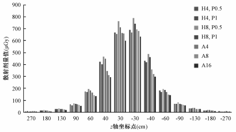
结果1. z轴各个测量点散射线剂量值:不论扫描参数如何,z轴方向上靠近扫描野中心散射线剂量值大,随着远离中心,散射线剂量逐渐减小,如图 1。

|
图 1 不同扫描方式、探测器宽度时z轴各个测量点的散射线剂量值 注:H为螺旋扫描;A为逐层扫描;4、8、16分别表示4、8、16 cm探测器宽度;P0.5、P1分别为螺距0.516 :1和0.984 :1 Figure 1 Scattered radiation dose at each measurement points on the z-axis when using different scanning modes and detector widths |
2. z轴两侧散射线剂量分布:不论探测器宽度大小,逐层扫描和螺旋扫描z轴方向人体头侧散射线剂量值均高于人体足侧散射线剂量值,见表 1。
|
|
表 1 不同扫描参数下z轴两侧的散射线比较 Table 1 Comparison of scattered radiation doses on both sides of z-axis under different scan parameters |
3.不同扫描参数下各点散射线剂量比较结果:逐层扫描时,不同探测器宽度的散射线分布均为探测器4 cm时最大,16 cm时最小(χ2=28.000,P < 0.05),其中坐标30 cm处散射线相差最大,其差值为67.5 μGy(测量值分别为665.50和598.00 μGy),相差11.29%。螺旋扫描时,不同探测器宽度的散射线分布均为探测器宽度8 cm时最大,4 cm时最小,螺距0.516 :1时(Z=-3.233,P < 0.05),坐标-30 cm处散射线相差最大,其差值为97.67 μGy(测量值分别为786.98和689.31 μGy),相差14.17%。螺距0.984 :1时(Z=-2.982,P < 0.05),坐标-30 cm处散射线相差最大,其差值为75.25 μGy(测量值分别为742.75和667.50 μGy),相差11.27%。螺旋扫描相同探测器宽度时,螺距为0.516 :1时高于螺距为0.984 :1的散射线,4 cm探测器宽度时(Z=-3.296,P < 0.05),坐标40 cm处散射线相差最大,其差值为22.86 μGy,(测量值分别为424.36和401.50 μGy),相差5.69%,8 cm探测器宽度时(Z=-3.296, P < 0.05),坐标30 cm处散射线相差最大,其差值为49.95 μGy,(测量值分别为762.95和713.00 μGy),相差7.01%。相应测量点差值见表 2。
|
|
表 2 不同扫描条件下同一测量点的对应差值(μGy) Table 2 Difference on the same measurement points under different scan parameters(μGy) |
讨论
随着CT设备不断更新换代、软硬件技术迅速发展,在临床检查中被大量应用,目前,CT扫描已成为最大的医源性人工辐射来源[7]。如何降低患者接受CT检查时的X射线辐射剂量,是目前普遍关注的问题。在临床工作中,应时刻遵循国际放射防护委员会(International Commission on Radiological Protection,ICRP)提出辐射防护最优化原则(as low as reasonably achievable,ALARA),即在满足临床诊断需求的前提下最大限度地降低辐射剂量[8]。
患者接受CT扫描时,除了受到主射线照射外,还受到机房内散射线的照射。此外对于脑出血、肺栓塞、主动脉夹层等危重症患者,常常需要家属留在机房内,儿童在接受CT检查时,也需家长在旁陪同,而对于需要行CT引导下穿刺的患者,其穿刺医生更是要近台操作。目前对于CT机房内散射线已有一些研究。白金城等[9]研究表明低剂量CT扫描可降低散射线强度,及散射线剂量与管电流、管电压有关,且曝光结束后5 s内机房散射线剂量最大。夏春潮等[10]测量并绘制了128层螺旋CT常规头部扫描时CT机房内的辐射场分布图,及水平面和立面的辐射场分布类似“8”字形。但对于现在所流行的高端宽体探测器CT不同探测器宽度对散射线的影响研究较少。本研究选用宽体探测器CT为研究对象,选取z轴方向为患者通常所受散射线的方向,探讨在不同扫描模式、不同探测器宽度时z轴的散射线分布规律,以此为临床防护提供依据。
研究结果显示逐层扫描时,4 cm探测器宽度散射线最大,分析原因可能为扫描长度均为16 cm,使用4 cm探测器宽度时X射线管需旋转4圈以完成扫描,将4个钟形曲线相互叠加,使用8 cm探测器宽度时X射线管需旋转2圈已完成扫描,使得每圈均有散射线叠加,从而导致所用探测器宽度越窄而散射线剂量越高。螺旋扫描长度同样为16 cm,当螺距相同时,8 cm探测器宽度的超范围扫描>4 cm探测器,故8 cm探测器宽度散射线最大。在探测器宽度和有效mAs均相同时,螺距小者其超范围扫描的剂量较大,所以螺距为0.516 :1的散射线高于0.984 :1的散射线。
本研究还有一些局限性:①本研究只对z轴方向上的辐射分布进行了测量,不能模拟整个CT室内的辐射场分布,可继续开展下一步实验予以完善。②扫描时所选管电压、管电流单一,不同的扫描参数可能会对辐射场的分布有一定影响。③本研究只考虑散射线剂量,而对图像质量考虑不足。不过有研究显示辐射剂量一致时,16 cm轴扫描模式的颅脑图像对比度噪声比(CNR)高于4 cm轴扫描模式、而噪声更低,且扫描速度缩短95%[11]。与本研究所示逐层扫描时尽量选用较宽探测器的结果一致。
综上所述,在使用宽体探测器CT时,在满足具体的临床需求的前提下,逐层扫描应尽量选择与扫描长度相当的宽探测器,螺旋扫描可适当选择窄探测器、大螺距,从而降低受检者、近台操作医务人员以及陪护人员的辐射剂量。
利益冲突 全体作者无利益冲突,进行该研究未接受任何不正当职务及财政获益,并对本研究的独立性和科学性予以保证作者贡献声明 郭森林负责研究设计、实验操作、结果统计分析、论文撰写;任悦负责协助实验操作及数据采集;牛延涛负责指导研究思路及论文修改
| [1] |
Brenner DJ, Hall EJ. Computed tomography-an increasing source of radiation exposure[J]. N Engl J Med, 2007, 357(22): 2277-2284. DOI:10.1056/NEJMra072149 |
| [2] |
Liu Z, Sun Y, Zhang Z, et al. Feasibility of Free-breathing CCTA using 256-MDCT[J]. Medicine (Baltimore), 2016, 95(27): e4096. DOI:10.1097/MD.0000000000004096 |
| [3] |
Greenberg SB. Dynamic pulmonary CT of children[J]. AJR Am J Roentgenol, 2012, 199(2): 435-440. DOI:10.2214/AJR.11.8014 |
| [4] |
Klink T, Nagel H, Schwartz B, et al. 256-MSCT image acquisition with sequential axial scans:evaluation of image quality and resolution in a phantom study[J]. Rofo, 2012, 184(3): 248-255. DOI:10.1055/s-0031-1299046 |
| [5] |
Norris JM, Kishikova L, Avadhanam VS, et al. Comparison of 640-Slice Multidetector Computed Tomography Versus 32-Slice MDCT for Imaging of the Osteo-odonto-keratoprosthesis Lamina[J]. Cornea, 2015, 34(8): 888-894. DOI:10.1097/ICO.0000000000000404 |
| [6] |
Shang D, Yue J, Li J, et al. Comparison of different width detector on the gross tumor volume delineation of the solitary pulmonary lesion[J]. J Cancer Res Ther, 2017, 13(4): 693-698. DOI:10.4103/jcrt.JCRT_1448_16 |
| [7] |
Sodickson A, Baeyens PF, Andriole KP, et al. Recurrent CT, cumulative radiation exposure, and associated radiation-induced cancer risks from CT of adults[J]. Radiology, 2009, 251(1): 175-184. DOI:10.1148/radiol.2511081296 |
| [8] |
Entrikin DW, Leipsic JA, Carr JJ. Optimization of radiation dose reduction in cardiac computed tomographic angiography[J]. Cardiol Rev, 2011, 19(4): 163-176. DOI:10.1097/CRD.0b013e31821daa8f |
| [9] |
白金城, 迟红卫, 王淑萍, 等. CT扫描中的散射线分析[J]. 中国中西医结合影像学杂志, 2017, 15(2): 177-179. Bai JC, Chi HW, Wang SP, et al. Analysis to the scattered radiation in CT scan[J]. Chin Imaging J Integr Tradit West Med, 2017, 15(2): 177-179. DOI:10.3969/j.issn.1672-0512.2017.02.016 |
| [10] |
夏春潮, 蒲进, 何玲, 等. 128层螺旋CT扫描的室内辐射场分布及辐射剂量[J]. 中华放射学杂志, 2016, 50(5): 388-390. Xia CX, Pu J, He L, et al. Radiation dose and radiation field distribution in 128-slice CT scanning[J]. Chin J Radiol, 2016, 50(5): 388-390. DOI:10.3760/cma.j.issn.1005-1201.2016.05.016 |
| [11] |
刘晓怡, 綦维维, 刘卓, 等. 头颅CT不同扫描方式的图像质量分析[J]. 中国医学影像学杂, 2017, 25(6): 418-421, 429. Liu XY, Qi WW, Liu Z, et al. Image quality assessment of brain CT witll different scaning modes[J]. Chin J Med Imaging, 2017, 25(6): 418-421, 429. DOI:10.3969/j.issn.1005-5185.2017.06.005 |
 2019, Vol. 39


