2. 510120 广州, 中山大学孙逸仙纪念医院神经科
2. Department of Neurology, Sun Yat-sen Memorial Hospital, Sun Yat-sen University, Guangzhou 510120, China
放疗在鼻咽癌(NPC)患者的治疗中起着至关重要的作用。然而,放疗的并发症,如放射性脑损伤[1]和放射性血管损伤[2]不可忽略。颈动脉的放射性动脉粥样硬化是头颈部放疗后临床相关的晚期并发症。与其他头颈部恶性肿瘤一样,鼻咽癌多种综合治疗后长期生存的改善导致放疗后患者颈动脉疾病发病率增加[3]。先前的研究发现,接受头部和颈部放疗的患者有更高的危险发生颈动脉狭窄[2, 4-5],最终患有短暂性脑缺血发作或中风。文献显示,严重狭窄是造成栓塞或缺血性中风的威胁[6-7]。随着放射性脑损伤的机制逐渐暴露,血管损伤已成为研究脑损伤关系的重点。对于放疗后的患者,放射性脑损伤患者与无脑损伤患者的脑血管和颈动脉损伤程度是否有差异?考虑到颞叶是鼻咽癌患者术后最脆弱的部位,常发生坏死,本研究探讨了NPC放疗后合并放射性颞叶坏死(temporal lobe necrosis,TLN)或无TLN患者的颈动脉内膜厚度(intima-media thickness,IMT)、颈动脉斑块形成及颈动脉和脑血流动力学差异,旨在揭示血管损伤与TLN的关系。
资料与方法1.入组标准、病例来源和分组:回顾性抽取2012年1月至2015年12月在本院收治的鼻咽癌患者作为本次研究对象。符合以下资格标准的患者进入本研究TLN组:鼻咽癌放射治疗史;无肿瘤复发、脑转移瘤、继发性颅内肿瘤、脑脓肿、脑梗死、脱髓鞘病、脑炎或其他中枢神经系统疾病的证据;MR扫描发现单侧或双侧TLN。TLN组患者共58例,其中男42例,女16例,年龄(38.2±5.1)岁。放疗后年限(4.58±1.89)年,化疗18(31.0%)例。33例鼻咽癌放疗后无TLN的患者进入本研究非TLN组,其中男24例,女9例,年龄(37.7±4.2)岁。放疗后年限(4.45±1.30)年,化疗11(33.3%)例。有以下任何一项的患者被排除在本研究之外:血管疾病的危险因素,包括高胆固醇血症、高血糖、高血压、糖尿病、吸烟或酗酒的历史;可能导致血管炎的疾病,包括结核病、梅毒和动脉炎;心血管疾病史。另外,选择29例同期在本院进行健康体检者作为对照组,男21例, 女8例, 年龄(38.1±5.4)岁。3组人群的人口统计学特征和非对照组间临床特征差异无统计学意义(P>0.05)。TLN患者常见症状为头痛(25例,43.1%),头晕(19例,32.8%),晕厥(17例,29.3%),偏瘫(16例,27.6%),记忆力减退(17例,29.3%)。
2.放射性颞叶坏死的诊断:TLN的诊断标准为T1加权图像上颞叶内表现为异常等信号或低信号,T2加权呈现为颞叶内异常高信号,T1增强扫描显示为颞叶内异常点状、指状、环状或不规则的强化病灶,强化病灶周围可伴或不伴水肿。排除肿瘤复发及其他疾病后可确诊[8]。
3.检查方法:所有扫描均由一位经验丰富的超声医师完成,使用LOGIC 700超声检查仪(美国GE公司)。扫描区域包括颈部两侧颈内动脉(ICAs)近端(即颈动脉分叉后1 cm内),测量ICAs近端内膜中层最厚处的厚度即为IMT值,还有ICAs检测区域内斑块的出现率。使用以色列RIMED公司的Trans-link9900经颅多普勒经颅多普勒(TCD)来采集基底动脉(BA)、双侧大脑中动脉(MCAs)和ICAs的血流速度数据。
4.统计学处理:使用SPSS 15.0软件进行分析。符合正态分布的数据用x±s表示。用U检验比较TLN组、非TLN组和对照组的临床特点、颈动脉内膜中层厚度、脑和颈动脉血流速度。采用χ2检验检测3组的颈内动脉斑块的发生情况,采用配对样本t检验比较两侧ICAs和MCAs的血管病变。利用Pearson相关性分析对放疗后时间、年龄与IMT的关系进行分析。P<0.05为差异有统计学意义。
结果1.颈动脉IMT的改变:各组颈动脉IMT如表 1所示,在58例TLN患者中,ICAs平均IMT明显大于33例放疗后无TLN者(t=10.208,P<0.05)。对照组与病例组(TLN组、非TLN组)ICAs的IMT差异均有统计学意义(t=18.624、8.221,P<0.05)。IMT与放疗后时限呈正相关(r=0.368,P=0.049),年龄与IMT无相关性(r=0.331,P=0.08)。
|
|
表 1 3组研究对象各动脉的检查情况(x±s) Table 1 Results of inspection in the three groups(x±s) |
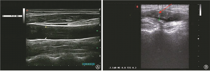
2.颈动脉斑块及狭窄情况:图 1显示了1例健康个体和1例TLN患者的颈动脉超声图像:其中40岁健康对照者,颈动脉管腔光滑,管径(d)=7.5 mm、IMT为0.64 mm;而38岁鼻咽癌放疗后TLN患者的颈动脉管腔狭窄(d=2.2 mm,IMT=7 mm),并出现粥样硬化斑块声像,管径狭窄率超过76%,箭头处显示为斑块。

|
图 1 健康对照者与TLN患者的颈动脉超声扫描影像图 A.健康对照者;B.TLN患者 注:箭头所指为斑块 Figure 1 Carotid ultrasonography in healthy individual and patient with TLN A. healthy individual; B. patient with TLN |
在放疗后的91例患者中,ICAs斑块总发生率为42.3%,明显高于对照组(χ2=17.886,P<0.05)。此外,TLN患者的斑块较无TLN差异有统计学意义(χ2=13.118,P<0.05)(表 1)。58例TLN患者的斑块面积明显大于33例放疗后无TLN者(t=4.086,P<0.05),两组均明显大于对照组(t=4.869、3.670,P<0.05)。以管腔狭窄率超过50%为界点,TLN患者与非TLN患者的动脉狭窄率差异无统计学意义(P>0.05),但均明显高于对照组(χ2=16.242、12.875,P<0.05)。
3. ICAs、MCAs、BA平均血流速度的改变:如表 1所示,TLN组ICAs的血流速度明显快于非TLN组(t=5.011,P<0.05),放射治疗后患者(TLN组或非TLN组)与健康者之间的差异均有统计学意义(t=9.074、3.194,P<0.05)。TLN组可观察到MCAs流速较非TLN组明显增快(t=5.035,P<0.05),两者均高于对照组(t=14.367、10.112,P<0.05)。TLN组、非TLN组和对照组的BA平均流速两两比较,差异均无统计学意义(P>0.05)。
4. TLN患者的ICAs、MCAs损伤的比较:在58例TLN患者中,28例出现单侧病变,30例出现双侧病变。其中28例单侧病变病例,病灶侧MCAs血流速度明显高于健侧(非病灶侧) [(79.48±10.79)cm/s vs.(68.07±13.67)cm/s,t=18.362,P<0.05],但病灶侧ICAs的IMT与非病灶侧差异无统计学意义[(2.34±0.57 mm)vs.(2.46±0.43)mm,P>0.05],斑块发生率差异也无统计学意义(46.4%、48.3%, P>0.05),ICAs血流速度也同样如此[(32.97±4.72)cm/s vs. (33.79±3.97)cm/s, P>0.05]。在30例双侧TLN患者中,两侧颈总动脉IMT、斑块发生率、MCAs流速及ICAs流速差异均无统计学意义(P>0.05)。
5.影响IMT、斑块发生率的单因素分析:在91例鼻咽癌放疗后的患者中,放疗疗程、放射剂量、病理分期与IMT增厚、斑块发生率有一定相关性(χ2=-17.635、12.006、-3.125,P<0.05),见表 2。
|
|
表 2 影响内膜中层厚度、斑块发生率的单因素分析比较(x±s) Table 2 ANOVA on IMT, incidence of plaques in ICAs(x±s) |
讨论
本研究评估了NPC放疗后TLN、无TLN患者和健康对照组的IMT、斑块形成和血流速度,并相互进行了对比分析。结果表明,放疗后患者的IMT、斑块形成发生率和血流速度与健康对照组之间存在显著差异。本研究结果支持放射治疗能导致血管损伤的认识。研究表明,颈总动脉、颈内动脉(CCAs/ICAs)是辐射后最脆弱的动脉[2]。辐射剂量[1, 9]和放射后时限[4, 10]是颈动脉狭窄的危险因素。一项前瞻性研究比较了放疗前和放疗后同年的同一患者的IMT值,发现放疗后第1年IMT显著增厚,24个月随访时IMT仍持续增厚[11]。在本研究中,3组之间的MCAs和ICAs血流速度有显著差异,BA则没有。BA不在辐射范围内可能是这种现象的原因。结果表明,放疗患者IMT增加,并且IMT与放射后时限呈正相关,这与以往研究一致[10-14]。辐照可直接损伤内皮,导致血小板粘附, 继而促进平滑肌细胞增殖和迁移[15],最终导致动脉壁增厚,甚至动脉狭窄或闭塞。已有实验证据支持内皮细胞是辐射损伤的主要靶细胞群体[16]。辐射诱导的血管狭窄病变具有特定的特征[17-18],仅限于受照射的区域。
鼻咽癌是剂量相关的肿瘤,在Ⅲ、Ⅳ期鼻咽癌的根治放疗中为达较高的控制率,鼻咽部的放疗必须达到较高的根治剂量,其局部控制率与局部的照射剂量相关。双侧颞叶下极的部位位于颅中窝,与海绵窦、破裂孔邻近, Ⅲ、Ⅳ期鼻咽癌可通过颅底解剖间隙侵及破裂孔、海绵窦等结构, 甚至可能侵及硬脑膜、颞叶, 射线在照射肿瘤及亚临床病灶时, 为能给予靶区较高剂量, 在放射治疗中颞叶不可避免会受到较高剂量的射线照射, 无论是采用传统二维放疗、三维适形放疗, 还是调强放射治疗, 放射性TLN均有较高的发生率[19]。本研究发现,放疗后的TLN患者与无TLN的患者在IMT、斑块形成的发生率以及MCAs和ICAs的血流速度有显著差异。结果表明,血管损伤的严重程度可能与TLN有关。有学者认为晚辐射对正常组织的迟发反应与各种主要细胞体系的损伤有关,血管内皮细胞的损伤在迟发性放射性组织损伤的发生发展中起主要作用[20]。然而到目前为止,没有直接证据显示大动脉损伤对辐射相关损伤的影响。血管的变化是晚期组织损伤的一个因素或仅仅是一个标志,这需要进一步研究。在本研究中,单侧TLN患者双侧动脉的对比分析显示,在ICAs的IMT、斑块形成方面没有显著差异,然而,病变同侧的MCAs的血流速度明显高于对侧,这表明颈动脉损伤与颞叶损伤并不平行。有研究发现,有中风或TIA症状患者的脑部血流量较无症状的明显减少,这与本研究在血流变方面的结果不同[21]。考虑到颞叶萎缩、囊变、液化和梗死可能与血管损伤有不同的表现,有待进一步研究。
利益冲突 所有作者未接受任何不正当的职务或财务利益作者贡献声明 叶剑虹负责设计研究方案、撰写论文等;陈庆瑜负责指导研究和审阅论文;容小明负责数据统计分析;吕瑞妍负责收集病例、数据采集
| [1] |
Lawrence YR, Li XA, El NI, et al. Radiation dose-volume effects in the brain[J]. Radiat Oncol Biol Phys, 2010, 76(3): S20-S27. DOI:10.1016/j.ijrobp.2009.02.091 |
| [2] |
Lam WW, Leung SF, So NM, et al. Incidence of carotid stenosis in nasopharyngeal carcinoma patients after radiotherapy[J]. Cancer, 2001, 92(9): 2357-2363. DOI:10.1002/1097-0142(20011101)92:9<2357::aid-cncr1583>3.0.co;2-k |
| [3] |
Thalhammer C, Husmann M, Glanzmann C, et al. Carotid artery disease after head and neck radiotherapy[J]. Vasa, 2015, 44(1): 23-30. DOI:10.1024/0301-1526/a000403 |
| [4] |
Cheng SW, Ting AC, Lam LK, et al. Carotid stenosis after radiotherapy for nasopharyngeal carcinoma[J]. Arch Otolaryngol Head Neck Surg, 2000, 126(4): 517-521. DOI:10.1001/archotol.126.4.517 |
| [5] |
Marcel M, Leys D, Mounier-Vehier F, et al. Clinical outcome in Patients with high-grade internal carotid artery stenosis after irradiation[J]. Neurology, 2005, 65(6): 959-961. DOI:10.1212/01.wnl.0000176033.64896.c6 |
| [6] |
Man BL, Fu YP, Chan YY, et al. Lesion patterns and stroke mechanisms in concurrent atherosclerosis of intracranial and extracranial vessels[J]. Stroke, 2009, 40(10): 3211-3215. DOI:10.1161/strokehah.109.557041 |
| [7] |
Thomas GN, Chen XY, Lin JW, et al. Middle cerebral artery stenosis increased the risk of vascular disease mortality among type 2 diabetic patients[J]. Cerebrovasc Dis, 2008, 25(3): 261-267. DOI:10.1159/000116303 |
| [8] |
任静, 周鹏, 刘锦, 等. 鼻咽癌放疗后迟发性放射性脑损伤的MRI特征[J]. 临床放射学杂志, 2009, 28(12): 1603-1606. Ren J, Zhou P, Liu J, et al. Imaging characteristics of MRI in delayed radiation induced brain injury after nasopharyngeal carcinoma radiotherapy[J]. J Clin Radiol, 2009, 28(12): 1603-1606. DOI:10.13437/j.cnki.jer.2009.12.045 |
| [9] |
Gianicolo ME, Gianicolo EA, Tramacere F, et al. Effects of external irradiation of the neck region on intima media thickness of the common carotid artery[J]. Cardiovasc Ultrasound, 2010, 8(1): 1-7. DOI:10.1186/1476-7120-8-8 |
| [10] |
Dorresteijn LD, KaPPelle AC, Scholz NM, et al. Increased carotid wall thickening after radiotherapy on the neck[J]. Eur J Cancer, 2005, 41(7): 1026-1030. DOI:10.1016/j.djca.2005.01.020 |
| [11] |
Muzaffar K, Collins SL, Labropoulos N, et al. A prospective study of the effects of irradiation on the carotid artery[J]. Laryngoscope, 2000, 110(11): 1811-1814. DOI:10.1097/00005537-200011000-00007 |
| [12] |
Chang YJ, Chang TC, Lee TH, et al. Predictors of carotid artery stenosis after radiotherapy for head and neck cancers[J]. Vasc Surg, 2009, 50(2): 280-285. DOI:10.1016/j.jvs.2009.01.033 |
| [13] |
Pereira LM, Biolo A, Foppa M, et al. A prospective, comparative study on the early effects of local and remote radiation therapy on carotid intima-media thickness and vascular cellular adhesion molecule-1 in patients with head and neck and prostate tumors[J]. Radiother Oncol, 2011, 101(3): 449-453. DOI:10.1016/j.radonc.2010.03.026 |
| [14] |
Li CS, Schminke U, Tan TY. Extracranial carotid artery disease in nasopharyngeal carcinoma patients with post-irradiation ischemic stroke[J]. Clin Neurol Neurosurg, 2010, 112(8): 682-686. DOI:10.1016/j.clineuro.2010.05.007 |
| [15] |
Tang Y, Luo D, Peng W, et al. Ocular ischemic syndrome secondary to carotid artery occlusion as a late complication of radiotherapy of nasopharyngeal carcinoma[J]. Neuroophthalmol, 2010, 30(4): 315-320. DOI:10.1097/wno.0b013e3181dee914 |
| [16] |
Abayomi OK. Neck irradiation, carotid injury and its consequences[J]. Oral Oncol, 2004, 40(9): 872-878. DOI:10.1016/j.oraloncology.2003.12.005 |
| [17] |
Pereira EB, Gemignani T, Sposito AC, et al. Low-density lipoprotein cholesterol and radiotherapy-induced carotid atherosclerosis in subjects with head and neck cancer[J]. Radiat Oncol, 2014, 11(9): 134. DOI:10.1186/1748-717X-9-134 |
| [18] |
Simonetti G, Pampana E, Di Poce I, et al. The role of radiotherapy in the carotid stenosis[J]. Ann Ital Chir, 2014, 85(6): 533-536. |
| [19] |
Zhou GQ, Yu XL, Chen M, et al. Radiation-induced temporal lobe injury for nasopharyngeal carcinoma:a comparison of intensity-modulated radiotherapy and conventional two-dimensional radiotherapy[J]. PLoS One, 2013, 8(7): e67488. DOI:10.1371/jouranal.pone.0067488 |
| [20] |
Lyubimova N, Hopewell JW. Experimental evidence to support the hypothesis that damage to vascular endothelium plays the primary role in the development of late radiation-induced CNS injury[J]. Br J Radiol, 2004, 77(918): 488-492. DOI:10.1259/bjr/15169876 |
| [21] |
Lam WW, Ho SS, Leung SF, et al. Cerebral blood flow measurement by color velocity imaging in radiation-induced carotid stenosis[J]. Ultrasound Med, 2003, 22(10): 1055-1060. DOI:10.7893/jum.2003.22.10.1055 |
 2018, Vol. 38


