Recently advanced technical changes in medical linear accelerators improved mechanical accuracy, stability and dosimetric control. Moreover, standard and add-on multi-leaf collimators based on conventional linear accelerators and special treatment units designed for stereotaxy or intensity modulated radiation therapy (IMRT) have been increasingly available. Developments in technology have increased the use of small fields for stereotaxy and the use of large fields composed of small fields [1-2].
A small field is defined as a field with a size smaller than the lateral range of charged particles. Dosimetry of small fields is more complicated than standard fields due to: ① the reference conditions specified in dosimetry protocols like the IAEA TRS-398[3]and AAPM TG-51[4], as beam calibration, cannot be realized in some treatment machines and ② dose measurement in small fields is not standard, such as lack of charged particle equilibrium (CPE), usual perturbing of detector to level of disequilibrium, source occlusion, high dose gradient, detector density, and detector volume averaging.
In order to develop a protocol of practice for small and non-standard fields, International Atomic Energy Agency (IAEA) in collaboration with American Association of Physicists in Medicine (AAPM) has provided a new formalism for the dosimetry of non-reference fields in 2008[5]. The new formalism defined a correction factor
An increasing number of detectors have been manufactured for small field dosimetry (eg, mini and micro ionization chambers, diodes, plastic scintillators, synthetic diamonds, radiographic and radiochromic films, polymer gels, etc.). The choice of a competent detector for the dosimetry of small photon fields requires well-established detector performance characteristics, for instance high reproducibility, sensitivity and linearity, minimum energy, angular, dose, and dose rate dependence, tissue equivalence, better spatial resolution[6].
There have been a growing number of authors reporting the dosimetric properties of different detectors. The corresponding correction factors
The aim of this paper is to study the dosimetric properties of PTW 60019 synthetic diamond detector for small photon beams in radiotherapy. The performances of such a synthetic diamond detector have been reported by various authors[19-22]. The performance of PTW 60019 diamond detector was compared with those of different reference detectors. In this work, experiments were performed to study its response sensitivity, linearity with dose, dose rate dependence and the suitability for measuring percent depth dose (PDD), off-axis ratio (OAR) and total scatter factor (Scp) in small photon fields. The performance of PTW 60019 was compared with those of a stereotactic field diode (SFD, IBA Dosimetry, Germany) and two ionization chambers: a PinPoint (PP, PTW 31016, PTW Freiburg, Germany) and a semiflex (PTW 31010, PTW Freiburg, Germany).
Materials and methods 1. Linear accelerator, electrometers and water phantomAll dosimetric measurements reported in this work were carried out with 6 and 10 MV photon beams generated by an Elekta Digital Linear accelerator (Elekta Crawley, UK). A Keithley electrometer (Model 6517B) was used for charge accumulation. An in-house motorized water phantom was used in this paper. It has a dimension of 30 cm× 30 cm× 30 cm. The polymethyl methacrylate (PMMA) has a 3.38 mm thick entrance window with water equivalent thickness of 3.89 mm. The other walls are made of 20 mm thick PMMA.
2. Diamond and reference detectorsThe synthetic single-crystal diamond detector tested in this work is PTW 60019 microDiamond detector (PTW Freiburg, Germany). The sensitive part of the diamond detector is a single crystal intrinsic diamond disk of 0.004 mm3, with diameter of 2.2 mm and thickness of 1 μm. This layer is sandwiched between a thin aluminum electrode and a conductive layer of P-type diamond doped with boron. The multilayered structure is mounted, through microwave-plasma-enhanced chemical vapor deposition (CVD), on a high-pressure high-temperature (HPHT) synthetic single crystal diamond. The device works as a Schottky barrier photodiode and without need of an external voltage bias. The effective point of measurement is 0.954 mm below the surface of the detector tip. Details on its detection mechanism and dosimetric properties can be found in literatures[23-25].
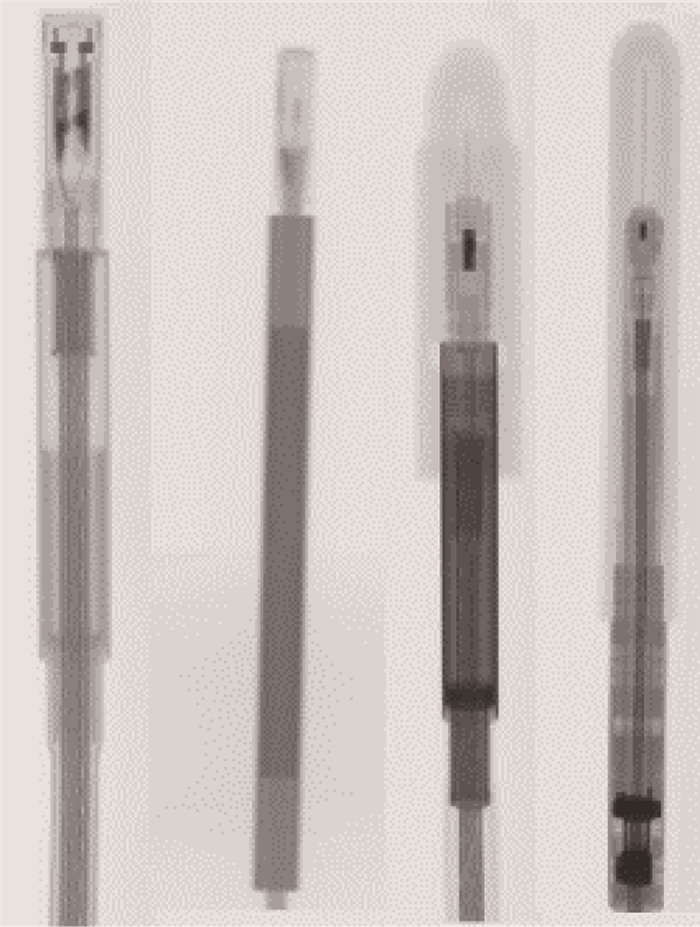
The dosimetric properties of PTW 60019 microDiamond detector are assessed by comparison with other three different types of commercial reference detectors: a stereotactic field diode (SFD, IBA Dosimetry, Germany), a PinPoint ionization chamber (PTW 31016, PTW Freiburg, Germany), and a semiflex ionization chamber (PTW 31010, PTW Freiburg, Germany), as shown in Figure 1.
|
Figure 1 Images of PTW 60019 microDiamond, IBASFD, PTW 31010 and PTW 31016 (from left to right) |
A stereotactic field diode IBA SFD operates at 0 V bias voltage. It has a sensitive volume of 0.017 mm3 with diameter of 0.6 mm and thickness of 60 μm. It was used as a reference detector for all dosimetric measurements.
A PinPoint ionization chamber PTW 31016 PinPoint was operated at 400 V bias voltage, with a sensitive volume of 0.016 mm3. The nominal cylindrical collection volume are 2.9 mm in diameter by 2.9 mm length. It was used as a reference detector for Scp measurements.
A semiflex ionization chamber PTW 31010 semiflex was operated at 400 V bias voltage, with a sensitive volume of 125 mm3 with diameter of 5.5 mm and thickness of 6.5 mm. It was used as a reference detector for dose rate dependence measurement and for PDD measurements for all field sizes.
For all measurements with PTW 60019 Diamond and IBA SFD, the detectors were oriented parallel to the central axis of the photon beam. Both PTW 31016 PinPoint and PTW 31010 semiflex were oriented with its axis perpendicular to the beam direction.
3. Dosimetric measurementsLinearity with dose, dose rate dependence, repeatability, PDD, OAR and Scp were studied, with all measurements being performed in an in-house water phantom. All fields were confined by jaws with multi-leaf collimator set to the same sizes.
Measurements of dose and dose rate dependence were performed using a 6 MV photon beam at 5 cm depth in water and in a 10 cm× 10 cm field with SSD of 100 cm. The linearity with dose was studied in the dose range from 100 to 1 000 MU. The repeatability was studied with associated experimental uncertainty in five consecutive measurements. The dose rate dependence of the detector was measured at dose rates of 37, 74, 150, 303 and 614 MU/min.
The measurement of PDD was conducted using 6 and 10 MV photon beams at a prescribed rate of 400 MU/min, in 1 cm× 1 cm, 2 cm × 2 cm, 3 cm × 3 cm, 5 cm × 5 cm and 10 cm × 10 cm fields. The SSD was set as 100 cm. An in-house developed software was used to control the detector position in the water phantom. Depth doses were measured at 2 mm steps from 6 mm to 150 mm for PTW 60019 Diamond and IBA SFD and from 8 mm to 150 mm for PTW 31010 semiflex.
The measurement of the OAR was carried out by using a 10 MV photon beam at 10 cm depth with SSD of 100 cm. The data were obtained for field sizes of 1 cm×1 cm, 2 cm×2 cm and 3 cm×3 cm.
The Scp were measured using 6 MV photon beams at 1.5 cm depth with SSD of 98.5 cm and using 10 MV at 2 cm depth with SSD of 98 cm. The field sizes used were 1 cm× 1 cm, 2 cm×2 cm, 3 cm×3 cm, 4 cm×4 cm, 5 cm×5 cm, 8 cm×8 cm and 10 cm×10 cm, respectively.
The reported experimental uncertainty refer to one standard deviation resulting from a consecutive set of five measurements
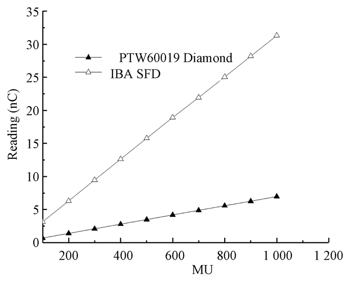
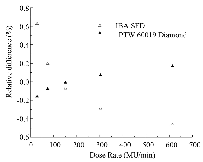
Results 1. Detector linearity with dose and dose rate dependenceThe linearity with dose in the 6 MV photon beam of the PTW 60019 Diamond and IBA SFD is shown in Figure 2. The R2 parameter of the linear best fit is 1 with a precision of 10-7. A deviation below 0.2% was observed over the entire range of investigated dose rates. A pre-irradiation of 1 Gy was performed before each measurement to verify the stability of PTW 60019 Diamond signal, with a variation of less than 0.2%. A sensitivity of 6.96 pC/MU is derived from the measured data as the slope of the linear fit. For IBA SFD, a maximum deviation from linearity of 0.8% and a sensitivity of 31.3 pC/MU are found. The dose rate dependence of PTW 60019 Diamond and IBA SFD relative to PTW 31010 semiflex is shown in Figure 3. A maximum variation obtained in the range of dose rates from 37 to 614 MU/min is 0.2% for PTW 60019 Diamond and 0.8% for IBA SFD, respectively. Thus, the PDD measured with the PTW 60019 Diamond will not require dose rate correction factor
|
Figure 2 Linearity with dose of PTW 60019 Diamond and IBA SFDdetectors as a function of absorbed dose to water at 5 cm depth |
|
Figure 3 Dose rate dependence of PTW 60019 Diamond and IBA SFDdetectors relative to the PTW 31010 semiflex response |
2. PDD measurements
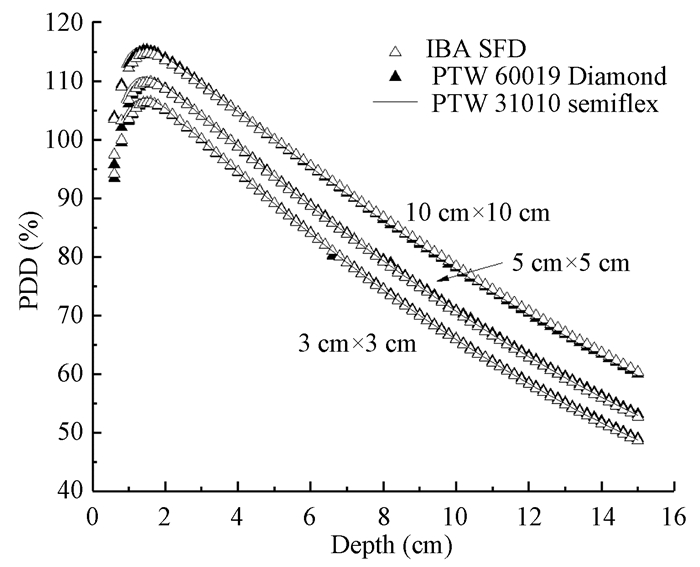
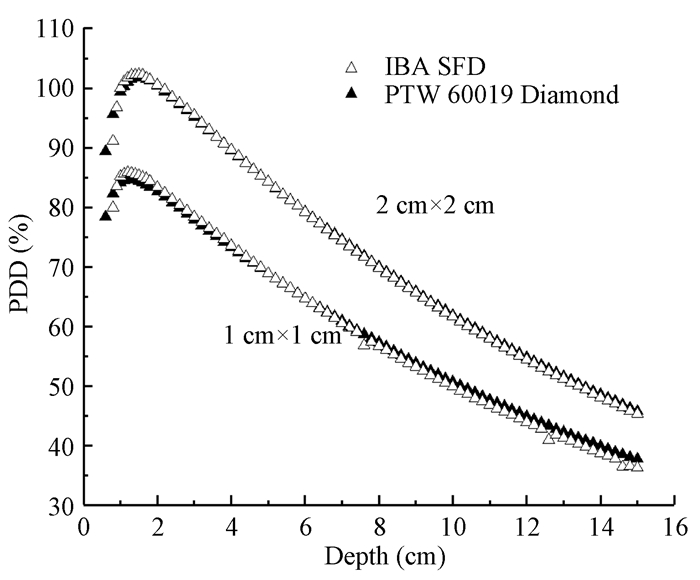
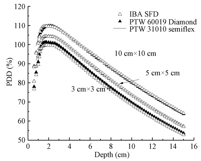
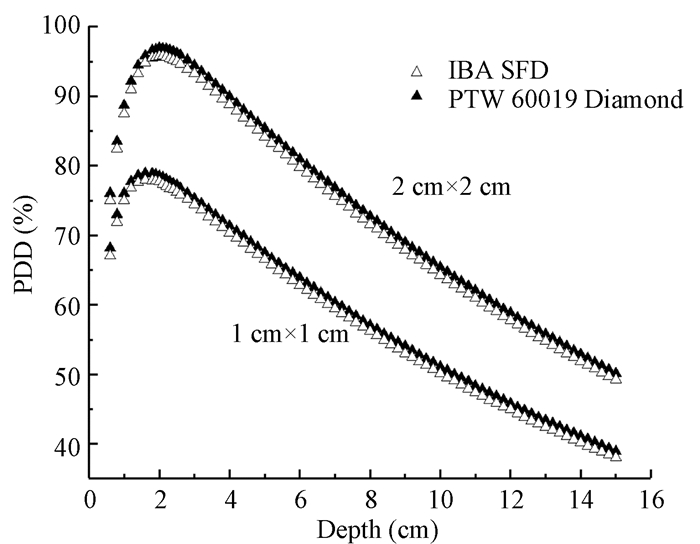
PDD measurements with different field sizes are displayed in Figure 4 and 5 for 6 MV photon beams and in Figure 6 and 7 for 10 MV photon beams. PDD curves are normalized to the a depth of 5 cm. In addition, the depths of maximum dose (dmax) positions, measured by the three detectors are shown in Table 1 for all field sizes investigated. Table 1 shows that PDD values measured by PTW 60019 Diamond were in good agreement with those by IBA SFD. The variations are higher in the smallest field size than in the 10 cm×10 cm field due to small error in the detector positioning in small beams. The PDD values measured by the PTW 31010 semiflex were slightly lower than the values measured by both PTW 60019 Diamond and IBA SFD because of the volume averaging effect.
|
Figure 4 PDDs measured in 6 MV photon beams with PTW60019 Diamond, IBA SFD and PTW 31010 semiflex for10 cm× 10 cm, 5 cm×5 cm and 3 cm×3 cm field sizes at SSD of 100 cm |
|
Figure 5 PDDs measured in 6 MV photon beams using PTW60019 Diamond, and IBA SFD for 2 cm×2 cm, 1 cm×1 cm field sizes at SSD of 100 cm |
|
Figure 6 PDDs measured in 10 MV photon beams using PTW60019 Diamond, IBA SFD and PTW 31010 semiflex for 10 cm×10 cm, 5 cm×5 cm and 3 cm×3 cm field sizes at SSD of 100 cm |
|
Figure 7 PDDs measured in 10 MV photon beams using PTW60019 Diamond, and IBA SFD for 2 cm×2 cm, 1 cm×1 cmfield sizes at SSD of 100 cm |
|
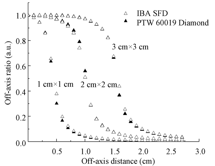
Figure 8 Half off-axis profiles measured in 10 MV photon beams ata depth of 10 cm by PTW 60019 Diamond and IBA SFD for 1 cm×1 cm, 2 cm×2 cm, 3 cm×3 cm field sizes at SSD of 100 cm |
|
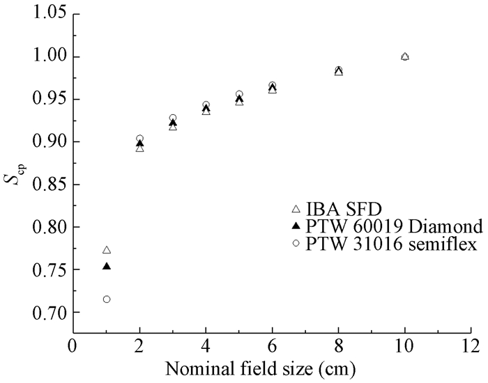
Figure 9 Total scatter factors measured with PTW 60019 Diamond, IBA SFD and PTW 31016 PinPoint in 6 MV photon beams at1.5 cm depth at a SSD of 98.5 cm |
|
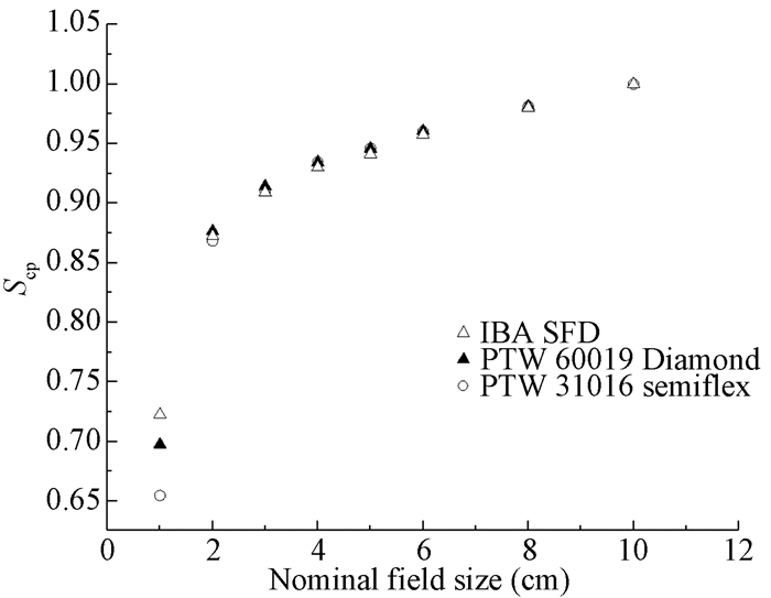
Figure 10 Total scatter factors measured with PTW 60019 Diamond, IBA SFD and PTW 31016 PinPoint in 10 MV photon beams at2 cm depth at a SSD of 98 cm |
|
|
Table 1 dmax measured with the PTW 60019 Diamond, the IBA SFD and the PTW 31010 semiflex, for 6, 10 MV photon beam and all the investigated field sizes(mm) |
PDD measurements with PTW 60019 Diamond and PTW 31010 semiflex were in a good agreement for the 10 cm×10 cm field under 6 MV photon beams, showing the differences less than 0.17% and a maximum difference of 0.51% for all depths greater than dmax. However, in the build-up region, the differences were 1.1% with a maximum difference of -2.5%.
A similar behavior can be observed for 5 cm×5 cm and 3 cm×3 cm field sizes. A slightly worse agreement is found between IBA SFD and PTW 31010 semiflex, with average differences of 0.38% and -1.65% at depths greater than dmaxand in the build-up region, respectively. For 2 cm×2 cm field size, the PDD curves measured by PTW 60019 Diamond were in agreement with those measured by IBA SFD, with a difference within 1%. PDD values for 1 cm×1 cm field size tend to be higher at depths greater than dmax, with a maximum difference 5.8%, due to a small error in the detector positioning under small photon beams, which could lead to an error of a few percent in PDD measurement.
For 10 MV photon beams, a very good agreement can be observed between the measurements by PTW 60019 Diamond, IBA SFD and PTW 31010 semiflex, down to the 3 cm× 3 cm field size, with the differences well below 1% at the depths greater than dmax and better than 2% in the build-up region. For both 1 cm×1 cm and 2 cm× 2 cm field sizes, the PDD curves measured by PTW 60019 Diamond were in agreement with those measured by the IBA SFD, with a difference within 1%.
3. Off axis profilesFigure 8 shows the off-axis profiles measured with PTW 60019 Diamond and IBA SFD for the 1 cm×1 cm, 2 cm×2 cm, 3 cm×3 cm field sizes at a depth of 10 cm. A good agreement between the profiles measured by the two detectors can be observed in the range of 1% to less. In the penumbra region, the differences tend to be higher with a maximum deviation of 25% in the 1 cm×1 cm field. Outside these high gradient regions, the differences are less than 10%.
4. Total scatter factorsThe Scp was measured for several field sizes ranging from 1 cm× 1 cm to 10 cm× 10 cm, respectively, for 6 MV beams at 1.5 cm, as in Figure 9, and for 10 MV beams at 2.0 cm depth, as in Figure 10. For field sizes larger than 1 cm×1 cm, both PTW 60019 Diamond and IBA SFD Scp values were in good agreement with those obtained with the PTW 31016 PinPoint, with a maximum deviation of 1%. For 1 cm×1 cm field size, the difference was up to 5% for PTW 60019 Diamond and 7% for IBA SFD. The Scp values obtained with PTW 60019 Diamond were higher than those obtained with IBA SFD, with a maximum difference of 2.5% for beam sizes down to 1 cm×1 cm. This behavior is attributed to the density effect of detection medium.
DiscussionIn the present work, we have evaluated the PTW 60019 Diamond detector for the dosimetry of small photon fields. The dosimetric properties of the microDiamond detector were assessed by comparing its PDD, off-axis profiles and total scatter factors with the those obtained by the reference detectors.
The detector was polarized at zero bias voltage. A good linear behavior with a deviation from linearity less than 0.2% was observed. The PTW 60019 Diamond has negligible dose rate dependence, with difference of about 0.2% in the dose rate range of 37 to 614 MU/min.
PDD curves and total scatter factors measured with the microDiamond were in good agreement with those obtained with the reference detectors. Spatial resolution was evaluated by means of off-axis profile measurements. The microDiamond presents a good spatial resolution for dose profile measurements, because of its small detection volume. It is a suitable candidate for small photon beam dosimetry.
Conflict of interest statement We declare that we do not have any commercial or associative interest that represents a conflict of interest in connection with the work submittedContribution statement of authors Chang Xue designed and carried out experiments, analyzed experimental results and drafted the manuscript; Wang Kun designed and carried outexperiments; Zhang Jian and Jin Sunjun carried out experiments; Wang Zhipeng carried out experiments and analyzed experimental results
| [1] | Das IJ, Morales J, Francescon P. Small field dosimetry:What have we learnt?[J]. Med Phys, 2016, 1417 (1): 060001 DOI:10.1063/1.4954111. |
| [2] | Das IJ, Ding GX, Ahnesjö A. Small fields:nonequilibrium radiation dosimetry[J]. Med Phys, 2008, 35 (1): 206-215. DOI:10.1118/1.2815356. |
| [3] | International Atomic Energy Agency. IAEA TRS 398. Absorbed dose determination in external beam radiotherapy[R]. Vienna: IAEA, 2000. |
| [4] | Almond PR, Biggs PJ, Coursey BM, et al. AAPM's TG-51 protocol for clinical reference dosimetry of high-energy photon and electron beams[J]. Med Phys, 1999, 26 (9): 1847-1870. DOI:10.1118/1.598691. |
| [5] | Alfonso R, Andreo P, Capote R, et al. A new formalism for reference dosimetry of small and nonstandard fields[J]. Med Phys, 2008, 35 (11): 5179-5186. DOI:10.1118/1.3005481. |
| [6] | Wilcox EE, Daskalov GM. Evaluation of GAFCHROMIC EBT film for CyberKnife dosimetry[J]. Med Phys, 2007, 34 (6): 1967-1974. DOI:10.1118/1.2734384. |
| [7] | Verhaegen F, Das IJ, Palmans H. Monte Carlo dosimetry study of a 6 MV stereotactic radiosurgery unit[J]. Phys Med Biol, 1998, 43 (10): 2755-2768. DOI:10.1088/0031-9155/43/10/006. |
| [8] | Benmakhlouf H, Johansson J, Paddick I, et al. Monte Carlo calculated and experimentally determined output correction factors for small field detectors in Leksell Gamma Knife Perfexion beams[J]. Phys Med Biol, 2015, 60 (10): 3959-3973. DOI:10.1088/0031-9155/60/10/3959. |
| [9] | Azangwe G, Grochowska P, Georg D, et al. Detector to detector corrections:a comprehensive experimental study of detector specific correction factors for beam output measurements for small radiotherapy beams[J]. Med Phys, 2014, 41 (7): 072103 DOI:10.1118/1.4883795. |
| [10] | Sterpin E, Hundertmark BT, Mackie TR, et al. Monte Carlo-based analytical model for small and variable fields delivered by TomoTherapy[J]. Radiother Oncol, 2010, 94 (2): 229-234. DOI:10.1016/j.radonc.2009.12.018. |
| [11] | Francescon P, Beddar S, Satariano N, et al. Variation of kQclin, Qmsr (fclin, fmsr) for the small-field dosimetric parameters percentage depth dose, tissue-maximum ratio, and off-axis ratio[J]. Med Phys, 2014, 41 (10): 101708 DOI:10.1118/1.4895978. |
| [12] | Bouchard H, Seuntjens J, Kawrakow I. A Monte Carlo method to evaluate the impact of positioning errors on detector response and quality correction factors in nonstandard beams[J]. Phys Med Biol, 2011, 56 (8): 2617-2634. DOI:10.1088/0031-9155/56/8/018. |
| [13] | Morin J, Beliveau-Nadeau D, Chung E, et al. A comparative study of small field total scatter factors and dose profiles using plastic scintillation detectors and other stereotactic dosimeters:the case of the CyberKnife[J]. Med Phys, 2013, 40 (1): 011719 DOI:10.1118/1.4772190. |
| [14] | Moignier C, Huet C, Makovicka L. Determination of the KQclinfclin, Qmsrfmsr correction factors for detectors used with an 800 MU/min CyberKnife© system equipped with fixed collimators and a study of detector response to small photon beams using a Monte Carlo method[J]. Med Phys, 2014, 41 (7): 071702 DOI:10.1118/1.4881098. |
| [15] | Francescon P, Cora S, Satariano N. Calculation of k(Q(clin), Q(msr)) (f(clin), f(msr)) for several small detectors and for two linear accelerators using Monte Carlo simulations[J]. Med Phys, 2011, 38 (12): 6513-6527. DOI:10.1118/1.3660770. |
| [16] | Benmakhlouf H, Sempau J, Andreo P. Output correction factors for nine small field detectors in 6 MV radiation therapy photon beams:a PENELOPE Monte Carlo study[J]. Med Phys, 2014, 41 (4): 041711 DOI:10.1118/1.4868695. |
| [17] | Bassinet C, Huet C, Derreumaux S, et al. Small fields output factors measurements and correction factors determination for several detectors for a CyberKnife© and linear accelerators equipped with microMLC and circular cones[J]. Med Phys, 2013, 40 (7): 071725 DOI:10.1118/1.4811139. |
| [18] | Morales JE, Crowe SB, Hill R, et al. Dosimetry of cone-defined stereotactic radiosurgery fields with a commercial synthetic diamond detector[J]. Med Phys, 2014, 41 (11): 111702 DOI:10.1118/1.4895827. |
| [19] | Underwood TS, Rowland BC, Ferrand R, et al. Application of the Exradin W1 scintillator to determine Ediode 60017 and microDiamond 60019 correction factors for relative dosimetry within small MV and FFF fields[J]. Phys Med Biol, 2015, 60 (17): 6669-6683. DOI:10.1088/0031-9155/60/17/6669. |
| [20] | Lárraga-Gutiérrez JM, Ballesteros-Zebadúa P, Rodríguez-Ponce M, et al. Properties of a commercial PTW-60019 synthetic diamond detector for the dosimetry of small radiotherapy beams[J]. Phys Med Biol, 2015, 60 (2): 905-924. DOI:10.1088/0031-9155/60/2/905. |
| [21] | Ciancaglioni I, Marinelli M, Milani E, et al. Dosimetric characterization of a synthetic single crystal diamond detector in clinical radiation therapy small photon beams[J]. Med Phys, 2012, 39 (7): 4493-4501. DOI:10.1118/1.4729739. |
| [22] | Laub WU, Crilly R. Clinical radiation therapy measurements with a new commercial synthetic single crystal diamond detector[J]. J ApplClin Med Phys, 2014, 15 (6): 4890 DOI:10.1120/jacmp.v15i6.4890. |
| [23] | Almaviva S, Marinelli M, Milani E, et al. Synthetic single crystal diamond diodes for radiotherapy dosimetry[J]. Nucl Instrum Methods Phys Res(Section A), 2008, 594 (2): 273-277. DOI:10.1016/j.nima.2008.06.028. |
| [24] | Almaviva S, Marinelli M, Milani E, et al. Chemical vapor deposition diamond based multilayered radiation detector:Physical analysis of detection properties[J]. J Appl Phys, 2010, 107 (1): 014511-014517. DOI:10.1063/1.3275501. |
| [25] | Betzel GT, Lansley SP, Baluti F, et al. Operating parameters of CVD diamond detectors for radiationdosimetry[J]. Nucl Instrum Methods Phys Res(Section.A), 2010, 614 (1): 130-136. DOI:10.1016/j.nima.2009.12.008. |
 2018, Vol. 38


