血管介入诊治是冠状动脉疾病诊治中一种安全有效的方法。至2020年,针对复杂冠状动脉疾病的介入治疗的数量将会达到现在的数倍[1]。血管介入手术中,手术人员需接近受检患者在长时间X射线透视环境下进行操作,相比其他手术,接受了较高的职业照射[2]和可能导致潜在的生物效应[3]。心血管介入手术人员,由于手术特殊性,职业照射量远高于其他血管介入手术人员。近年来,过量辐射风险已越来越引起了人们的重视和关注。剂量面积乘积(dose-area product,DAP)是测量仪器置于X射线管和患者体表之间,对通过的射线束进行测量所得数据(DAP=剂量×测量仪器面积),在临床职业照射环境研究中,许多研究针对DAP值与手术人员的辐射剂量之间联系做出了大量的论证[4],认为DAP值可作为介入手术辐射剂量评估的重要证据,并已被临床广泛接受和采用。本研究通过DAP数据库相关数据,对辐射风险做出早期预判和快速干预,从而实现降低临床介入人员辐射风险的目的。
资料与方法1.一般资料:回顾性分析2016年4月1日至6月30日院内心血管介入检查1 858例病例。数据库字段分别包括:检查日期、患者ID号、性别、年龄、身高、体重、手术类型(冠状动脉造影术、经皮冠状动脉介入治疗术、先天性心脏病治疗术、永久性起搏器植入术)、手术剂量参数(透视时间、透视DAP值、总DAP值)、数字减影血管造影(DSA)设备型号、手术主治医师、助手医师、配合护士,数据记录完全在Microsoft Office Excel表格中完成。
2.成像系统及方法:所有DAP数据均实时测自德国西门子AXIOM Artis FC和AXIOM Artis Zee Biplane数字减影血管造影(DSA)数字成像设备,分别为影像增强器(image intensifier,Ⅱ)和平板探测器(flat panel detector,FPD)成像系统。采集参数:透视与电影采集帧频均为15帧/s,自动曝光采集。数据信息来自术后设备文件检查协议,包括透视时间、透视DAP(Artis FC无此数据)和总DAP。根据设备成像系统分组,Ⅱ组1 242例病例,其中冠状动脉造影术(CAG) 589例,经皮冠状动脉介入治疗术(PCI) 521例;FPD组616例病例,其中CAG 327例,PCI 250例。分别行相关资料和同类手术数据比较与分析。
3.统计学处理:计量资料以x±s表示。采用SPSS 19.0软件进行分析。经正态性检验符合正态分布后应用独立样本t检验对成像设备分组CAG和PCI样本的体质量指数(BMI)、透视时间和DAP数据进行比较。P < 0.05为差异有统计学意义。
结果1. PCI及其他手术在不同成像设备的透视时间和DAP值:结果列于表 1。表 1显示Ⅱ成像系统累积病例数、透视时间均明显高于FPD成像系统,DAP值接近相等。FPD成像系统PCI透视剂量是电影采集剂量(PCI总DAP-透视DAP)的2.8倍。PCI总DAP值达9 517 926 μGy ·m2,占总样本DAP值的77%。
|
|
表 1 不同成像设备上冠状动脉治疗术和其他手术透视时间、透视剂量面积乘积值和总剂量面积乘积值比较 Table 1 Comparison of FT, fluoroscopy DAP and total DAP values between PCI and other procedures using different angiography systems (Ⅱ and FPD) |
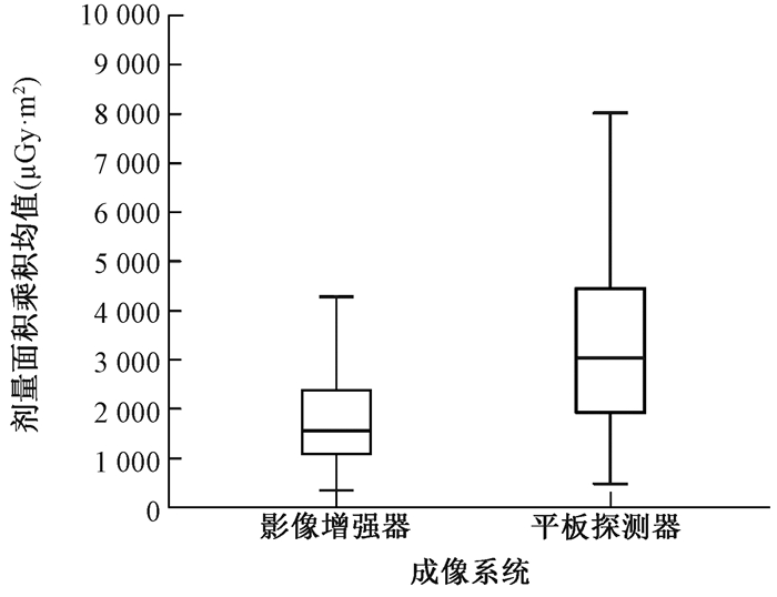
2.两种成像设备参数比较:结果列于表 2。由表 2可知,各组BMI差异均无统计学意义(P>0.05), Ⅱ和FPD成像设备CAG透视时间差异无统计学意义(P>0.05),PCI透视时间差异有统计学意义(t=-3.634,P < 0.05),CAG和PCI的DAP差异均有统计学意义(t=-10.664、11.239,P < 0.05)。FPD设备CAG检查DAP均值(3 718 μGy ·m2)高于Ⅱ设备(1 938 μGy ·m2),见图 1。
|
|
表 2 Ⅱ和FDP设备上冠状动脉造影和冠状动脉治疗术手术例数、体质量指数、透视时间和剂量面积乘积比较(x±s) Table 2 Case numbers, BMI values, FT and DAP values in CAG and PCI for Ⅱ and FPD systems(x±s) |

|
图 1 影像增强器(左)与平板探测器(右)成像设备行CAG检查中采集剂量面积乘积均值箱形图 Figure 1 Box-and-whisker plots for mean DAP values for CAG on the left Ⅱ system and right FPD system |
讨论
DSA设备均具备DAP采集功能,DAP是累积剂量与测量仪照射面积的乘积,不受X射线管与测量仪间的距离的变化而改变,对于术中需采用多方位、多角度采集的心血管介入手术,DAP监测有一定的优势,已被临床广泛作为辐射剂量监测依据。DAP数据库是便捷的获得辐射剂量证据的方法。
本研究Ⅱ组总透视时间高于FPD组75%,总DAP值低于FPD组,鉴于DAP值与透视时间有较好的正相关性[5],推测设备之间辐射剂量存在明显差异。不同成像设备同类资料比较,排除病例BMI干扰因素(DAP值与受检者BMI有正相关性),发现CAG透视时间差异较小,分析与检查技术要求相对简单和成熟有关;PCI透视时间存在差异主要受病变复杂、操作难度和术者技巧影响;接受CAG研究结果更具客观性。CAG结果显示,Ⅱ成像系统DAP值明显低于FPD成像系统,与Wiesinger等[6]报道相符。同时,FPD透视时间略少于Ⅱ,证明FPD设备的DAP值采集高于Ⅱ设备。从Biplane(FPD)成像系统的资料中提取BMI均值(27.1±1.3) kg/m2和透视时间(2.8±2.3) min的137例CAG样本,所获得的DAP数据为(4 157±2 698)μGy·m2,明显高于Kastrati等[7]报道西门子Biplane成像系统431例CAG样本,BMI(28.3±5.3) kg/m2,透视时间(4.6±4.3) min,DAP数据(3 193±2 358)μGy·m2。从而推断FPD成像系统采集剂量较高。对Ⅱ组剂量明显低于FPD组给出了充分的客观数据和确切的理论依据。本研究发现,FPD设备上PCI透视总DAP值明显高于造影,PCI总DAP值占全部样本总DAP值的77%,表明心血管介入中辐射剂量的来源主要是PCI。推测降低PCI手术透视时间是降低心血管介入手术辐射风险的重要手段。
应对策略主要有:联系设备工程师,对FPD成像参数进行调整;满足临床手术信息前提下,降低手术辐射剂量;暂时减少Biplane成像系统手术安排,对已安排手术采用改变采集协议,建议FPD成像系统造影采集帧频由15帧/s改变为10帧/s,透视帧频则从15帧/s降至7.5帧/s,尤其在PCI手术中,这将有效降低操作人员30%和受检患者19%的辐射剂量[8],积极推荐术中透视序列储存替代电影采集(Artis FC无此功能)和尽量缩小缩光器的操作方法[9]。
在职业辐射防护中,DAP数据库具备采集便捷、数据详细和随时分析的优点,可及时发现辐射剂量差异和来源并采取干预,相比剂量计Hp (10) 的信息量和直读式剂量计[10]的成本有一定优势,为临床辐射防护和个人健康及剂量管理制度实施提供了有效、细化的数据,是临床介入应用中值得推广的辐射防护优化工具。不足之处包括:录入数据为手工输入,存在耗时和差错,需要反复纠正和确认。数据来自一个导管室,样本来源局限。因此,及早设计出DAP数据库相匹配的采集协议数据同步存储和分析软件,建立数据库的互联网共享,将为职业辐射风险防护的大数据分析提供更加快捷准确的宏观数据。
利益冲突 作者无利益冲突,排名无争议,未接受任何不当职务或财务利益作者贡献声明 丁海岭负责本论文的撰写;王永春、江薇参与数据收集和分析处理
| [1] | Farajollahi A, Rahimi A, Khayati SE, et al. Patient's adiation exposure in coronary angiography and angioplasty:The impact of different projections[J]. J Cardiovasc Thorac Res, 2014, 6 (4): 247-252. DOI:10.15171/jcvtr.2014.020. |
| [2] | Taghi M, Toossi B, Mehrpouyan M, et al. Preliminary results of an attempt to predict over apron occupational exposure of cardiologists from cardiac fluoroscopy procedures based on DAP (dose area product) values[J]. Australas Phys Eng Sci Med, 2015, 38 (1): 83-91. DOI:10.1007/s13246-014-0326-1. |
| [3] | Jolly SS, Cairns J, Niemela K, et al. Effect of radial versus femoral access on radiation dose and the importance of procedural volume:a substudy of the multicenter randomized rival trial[J]. JACC Cardiovasc Interv, 2013, 6 (3): 258-266. DOI:10.1016/j.jcin.2012.10.016. |
| [4] | Bor D, Olgar T, Onal E, et al. Assessment of radiation doses to cardiologists during interventional examinations[J]. Med Phys, 2009, 36 (8): 3730-3736. DOI:10.1118/1.3168971. |
| [5] |
冯俊, 王爱玲, 程景林, 等. 不同类型心血管介人手术辐射剂量分析[J].
中华放射医学与防护杂志, 2012, 32 (4): 416-419. Feng J, Wang AL, Cheng JL, et al. Analysis of X-ray radiation doses from different types of intervention for cardiovascular patients[J]. Chin J Radiol Med Prot, 2012, 32 (4): 416-419. DOI:10.3760/cma.j.issn.0254-5098.2012.04.024. |
| [6] | Wiesinger B, Kirchner S, Blumenstock G, et al. Difference in dose area product between analog image intensifier and digital flat panel detector in peripheral angiography and the effect of BMI[J]. Rofo, 2013, 185 (2): 153-159. DOI:10.1055/s-0032-1330276. |
| [7] | Kastrati M, Langenbrink L, Piatkowski M, et al. Reducing radiation dose in coronary angiography and angioplasty using image noise reduction technology[J]. Am J Cardiol, 2016, 118 (3): 353-356. DOI:10.1016/j.amjcard.2016.05.011. |
| [8] | Abdelaal E, Plourde G, MacHaalany J, et al. Effectiveness of low rate fluoroscopy at reducing operator and patient radiation dose during transradial coronary angiography and interventions[J]. JACC Cardiovasc Interv, 2014, 7 (5): 567-574. DOI:10.1016/j.jcin.2014.02.005. |
| [9] | Seiffert M, Ojeda F, llerleile KM, et al. Reducing radiation exposure during invasive coronary angiography and percutaneous coronary interventions implementing a simple four-step protocol[J]. Clin Res Cardiol, 2015, 104 (6): 500-506. DOI:10.1007/s00392-015-0814-7. |
| [10] |
黄卓, 范瑶华, 李文炎, 等. 直读式剂量计用于介入职业人员眼晶状体剂量实时监测方法的研究[J].
中华放射医学与防护杂志, 2016, 36 (12): 926-934. Huang Z, Fan YH, Li WY, et al. Study of real-time measurements of occupational staff's eye lens doses by direct-reading dosimeters in interventional procedures[J]. Chin J Radiol Med Prot, 2016, 36 (12): 926-934. DOI:10.3760/cma.j.issn.0254-5098.2016.12.011. |
 2017, Vol. 37


