放射治疗是肿瘤常用的治疗方式,近年来对肿瘤照射的精度要求越来越高,肿瘤存在治疗过程中的变化和分次治疗间的位移误差,如呼吸和蠕动运动、日常摆位误差、靶区收缩等引起放疗剂量分布的变化和对治疗计划的影响等方面的情况[1-2]。需要利用锥形束CT (cone beam computed tomography,CBCT) 在治疗过程中对肿瘤及正常器官进行实时监控,将获取的CBCT图像和CT图像进行对比,以观察患者肿瘤位置的变化。目前,图像引导放疗 (image guide radiation therapy,IGRT)[3-4]和自适应放疗 (adaptive radiation therapy,ART)[5]的使用越来越广泛,为了对比患者定位CT图片与上述实时获取的CBCT图像的差异,需要对患者的CT和CBCT图像进行配准[6-7]。配准有很多种算法[8-9],如:基于外部特征、基于内部和基于像素密度的配准算法等。图像分割与配准工具包 (insight segmentation and registration toolkit, ITK) 是一个用C++语言建立的面向对象采用模板编程技术的、跨平台的开源软件开发包[10]。本研究采用基于灰度均方差测度的ITK程序包,分析CBCT与CT配准范围对配准精度的影响。
材料与方法1.扫描设备与图像参数:CT图像由日本东芝医疗系统公司生产的Aquilion PRIME型CT扫描得到,层厚5 mm,管电压为120 kV,视野 (FOV) 为50 cm,管电流为150 mA (头部和胸部)、175 mA (腹部),像素为512×512。CBCT图像由美国瓦里安公司生产的Ⅸ型加速器扫描得到,层厚2.5 mm,管电压为110 kV,FOV为45 cm,管电流为20 mA,像素为512×512。在图像配准前,都已将CT和CBCT二者的图像重建为1 mm相同的层厚。
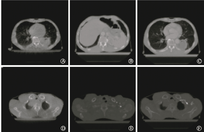
2.CT与CBCT图像:对来自南京医科大学附属常州市第二人民医院进行头部、胸部、腹部影像学检查的患者各5例行定位CT扫描,前3次摆位行CBCT扫描。将扫描后的CT和CBCT图像用MATLAB R2009a进行配准范围的处理,共4种模式,见图 1。模式1为临床上患者CT扫描长度双侧均大于CBCT的扫描长度,即CT的配准范围大于CBCT的配准范围;模式2在模式1的基础上将CT的配准范围双侧调整扫描至与CBCT相同,此时的CBCT和CT配准范围相同;模式3在模式2的基础上保持CBCT不动,将CT扫描的首末层平移5 cm,模式3可代表临床上扫描的CT图像中心偏离肿瘤中心的情况,CBCT扫描时图像中心通常都是肿瘤中心,在肿瘤中心两侧层数是一致的,若CT扫描偏离肿瘤中心太远,很有可能造成肿瘤一侧CT层数较多,另一侧CT层数较少,甚至刚好在肿瘤的边缘;模式4在模式2的基础上将CBCT和CT调整至两边同时减少2 cm。模式1和模式2配准所对应的DICOM图像见图 2。AB之间的图像对应于图 1中模式1的胸部CBCT图像,CD之间的图像对应于图 1中模式1的胸部CT图像,EF之间的图像对应于图 1中模式2的胸部CT图像。AB与CD配准即为图 1中胸部模式1配准,AB与EF配准即为图 1中胸部模式2配准。模式3和模式4对应的DICOM图像是基于模式2进行的平移和内缩,此处不赘述。

|
图 1 CBCT和CT配准的不同模式 Figure 1 Different modes of CBCT and CT registration |

|
图 2 同一患者的胸部DICOM图像 A.模式1胸部CBCT图像的第一层;B.模式1胸部CBCT图像的最后一层;C.模式1胸部CT图像的第一层;D.模式1胸部CT图像的最后一层;E.模式2胸部CT图像的第一层;F.模式2胸部CT图像的最后一层 Figure 2 The DICOM images of chest for one same patient A.The first layer CBCT image of chest in mode 1; B.The last layer CBCT image of chest in mode 1; C.The first layer CT image of chest in mode 1; D.The last layer CT image of chest in mode 1; E.The first layer CT image of chest in mode 2; F.The last layer CT image of chest in mode 2 |
3.CT与CBCT图像配准:将4种不同模式DICOM格式的CT和CBCT图像先用格式转换程序转化为short型Raw数据格式,再将short型Raw数据格式转化为float型Raw数据格式,以便后续进行配准。用Cmake软件将转换格式程序的代码进行编译,设置源代码目录以及编译目录,然后在VS2012开发平台上将代码编译成可执行程序,最后将float型Raw数据格式的CBCT和对应的CT图像用ITK工具包中的配准程序进行三维配准,配准的程序也需要用Cmake软件将配准代码进行编译,再编译成可执行程序。为检验配准的重复性,本研究每个患者每种模式的配准均重复5次实验。配准参数:优化器为常规步长梯度下降优化器,迭代次数为200,最小步长为0.001,测度类型为基于灰度的平均方差 (mean square difference,MSD) 测度,对4种不同模式的配准结果进行比较分析。
4. MSD算法:MSD是关于灰度的函数,反映了CBCT和CT两幅图像差异的大小,两幅图像差异越大,则MSD的值越大,若差异越小,则MSD的值越小。MSD的函数表达式为:
| $ {\rm{MSD}} = \frac{{\sum\limits_0^X {{{({F_i}-{M_i})}^2}} {\rm{ }}}}{X} $ | (1) |
式中,Fi和Mi分别为其假设在参考图像 (fixed image)F和浮动图像 (moving image)M上的i处的灰度值,X为像素点的个数。
5.统计学处理:采用SPSS 19.0软件进行数据分析。对模式2的腹部、头部、胸部的MSD测度值与其他3种模式进行正态性检验,符合正态分布,行配对t检验。P < 0.05为差异有统计学意义。
结果本研究对腹部、头部、胸部各5位患者的图像配准后得到的MSD值结果和配对t检验结果见表 1。从MSD值结果可以看出,模式3配准的MSD值最高,其次是模式1,模式2和模式4的MSD值最小且几乎相等 (P>0.05)。模式3的MSD值最高表明当扫描的CT图像中心偏离肿瘤中心较远时,配准精度最低。模式2与模式1、3比较,差异有统计学意义 (t=-19.601~-4.164,P < 0.05)。
|
|
表 1 患者4种模式配准的平均方差值比较 Table 1 The MSD values for four modes |
将CT图像作为参考图像,CBCT图像作为浮动图像,采用ITK包中的配准程序进行配准,同一患者腹部的模式1和模式2配准结果任意一层见图 3。如图所示,D是模式1配准结果中与A、B对应的任意一层,E是模式2配准结果中与A、C对应的任意一层。D的配准结果如图所示,明显不如E的配准结果理想,表明模式2的配准效果优于模式1配准结果。图 3仅为模式1和模式2配准结果,模式3和模式4的配准结果分别与模式1和模式2配准结果类似,此处不再赘述。

|
图 3 同一位患者腹部模式1和模式2配准结果 A.为模式1腹部任意一层CBCT图像;B.为模式1与A对应的同一层腹部CT图像;C.为模式2与A对应的同一层腹部CT图像;D.为模式1配准结果;E.为模式2配准结果 Figure 3 The registration results of mode 1and mode 2 on one patient′s abdomen A.Any CBCT image of abdomen in mode 1; B.The same layer of abdomen CT images corresponding to A in mode 1; C.The same layer of abdomen CT images corresponding to A in mode 2; D.The registration result of mode1; E.The registration result of mode 2 |
讨论
随着放疗技术的发展,对精准放射治疗剂量照射的精度要求越来越高,治疗过程中图像配准的技术研究也日益重要[11]。由于ITK性能稳定和开源的特点,其越来越多地应用在医学图像处理领域[12-15]。Godley等[16]将ITK应用于对自适应放疗中前列腺区域进行形变配准,并认为ITK配准后的结果可有效地校正分次治疗引起的肿瘤位置变化。Liu等[17]应用ITK对刚体配准与非刚体配准结果进行分析,结果表明非刚体配准结果优于刚体配准结果。Museyko等[18]将ITK应用于骨组织的配准。有学者指出,图像引导中的图像范围选取会影响配准精度[19-20]。彭应林等[21]在CBCT引导摆位中,指出仅对靶区附近范围进行图像配准,配准精度差于配准患侧和配准体廓组,并认为选择灰度配准算法以及采用配准患侧方式进行图像配准能够获得临床要求的较好的精度和较高的效率。
本研究结果显示,4种配准模式中,模式3的配准精度最差,其次是模式1配准,模式2和模式4配准精度最好。模式2和模式4比较的意义是CBCT和CT配准范围保持相同的前提下,在同一解剖结构上增大和缩小一定的配准范围不影响配准的精度。基于均方差测度的图像配准过程是对配准范围内的参考图像和浮动图像所有像素点的特征进行相似性比较的数学最优化搜索,配准运算结果与配准特征的数量和分布相关,因此对配准范围及相对位置非常敏感,配准范围内的组织结构形状和不均匀性等都会对配准效果产生影响,从而影响到参考图像和浮动图像配准的精度。当选择的配准范围不同时,实际上相当于对不同组织结构形状进行配准运算得到的最优解,其结果就有可能并非最优值,这也会影响配准的精度。孙文泽等[22]对中心型小细胞肺肿瘤与肿瘤+椎体两种配准范围进行了研究,相当于在原肿瘤基础上增加椎体扩大配准范围,认为配准范围会影响到配准精度。本研究不仅在原基础上扩大靶区配准范围,而且在原基础上对CT扫描图像的中心偏离肿瘤中心较远的情况进行研究分析。CBCT和CT配准时,相同的配准范围能显著提高配准精度。
综上,针对参考图像和浮动图像的配准范围对精度的影响,本研究利用均方差测度算法对4种模式的配准范围进行配准。实验结果证明,参考图像和浮动图像的配准范围越接近,配准精度越高,在实际配准时,配准范围应予考虑,在图像配准之前应尽量使参考图像和浮动图像的配准范围相同,以保证配准结果的精度。在算法实现过程中发现,虽然本研究采用的计算机平台配置较高,但每个患者每种模式配准计算时间一般要10~30 min不等,耗费大量的时间。另外,本研究对CBCT与CT图像的配准是基于ITK平台运算的三维静态配准,肿瘤在治疗的过程中是实时运动的。因此,针对本研究配准效率较慢的缺陷,以后将会对医学图像配准不同算法进行更深入的研究,以提升配准的精度和效率;针对肿瘤运动的情况,将来会对基于ITK平台运算的4维动态配准进行更深入研究。
利益冲突 本研究还接受江苏省常州市高层次卫生人才培养工程 (2016CZLJ004) 和江苏省常州市社会发展项目 (CJ20160029) 资助。本人与本人家属、其他研究者,未因进行该研究而接受任何不正当的职务或财务利益,在此对研究的独立性和科学性予以保证作者贡献声明 眭建锋设计研究方案,收集数据后统计并起草论文;孙鸿飞和高留刚协助提供符合入组病例;倪昕晔指导、监督试验进行,修改论文;谢凯和林涛负责进行试验及数据的初步处理
| [1] |
吴爱东, 张绍虎, 张红雁, 等. 锥形束CT测量胸段食管癌调强放疗摆位误差剂量学的影响[J].
中华放射医学与防护杂志, 2012, 32 (4): 379-382. Wu AD, Zhang SH, Zhang HY, et al. A kV cone-beam CT based analysis of thesetup errors and the corresponding impact on the dose distribution of intensity modulated radiotherapy for thoracic esophageal carcinoma[J]. Chin J Radiol Med Prot, 2012, 32 (4): 379-382. DOI:10.3760/cma.j.issn.0254-5098.2012.04.011. |
| [2] |
徐子海, 伍锐, 朱超华, 等. 肿瘤靶区呼吸运动位移误差补偿系统模型的研究[J].
生物医学工程研究, 2010, 29 (2): 133-135. Xu ZH, Wu R, Zhu CH, et al. The model development for compensating the displacement error of tumor target breathing movement[J]. Biomed Eng Res, 2010, 29 (2): 133-135. DOI:10.3969/j.issn.1672-6278.2010.02.015. |
| [3] | Kanakavelu N, Samuel EJ. Accuracy in automatic image registration between MV cone beam computed tomography and planning kV computed tomography in image guided radiotherapy[J]. Rep Pract Oncol Radiother, 2016, 21 (5): 487-494. DOI:10.1016/j.rpor.2016.07.001. |
| [4] | Ward MC, Ross RB, Koyfman SA, et al. Modern image-guidedintensity-modulated radiotherapy for oropharynx cancer and severe latetoxic effects: implications for clinical trial design[J]. JAMA Otolaryngol Head Neck Surg, 2016, 142 (12): 1164-1170. DOI:10.1001/jamaoto.2016.1876. |
| [5] | Brown E, Owen R, Harden F, et al. Head andneck adaptive radiotherapy: predicting the time to replan[J]. Asia Pac J Clin Oncol, 2016, 12 (4): 460-467. DOI:10.1111/ajco.12516. |
| [6] | Foley D, O'Brien DJ, León-Vintró L, et al. Phase correlation applied to the 3D registration of CT and CBCT image volumes[J]. Phys Med, 2016, 32 (4): 618-624. DOI:10.1016/j.ejmp.2016.02.009. |
| [7] | Li X, Zhang YY, Shi YH, et al. Evaluation of deformable image registration for contour propagation between CT and cone-beam CT images in adaptive head and neck radiotherapy[J]. Technol Health Care, 2016, 24 (Suppl 2): S747-S755. DOI:10.3233/THC-161204. |
| [8] | Klein S, Starting M, Murphy K, et al. Elastix: a toolbox for intensity-based medical image registration[J]. IEEE Trans Med Imaging, 2010, 29 (1): 196-205. DOI:10.1109/TMI.2009.2035616. |
| [9] | Qu J, Gong L, Yang L. A 3D point matching algorithm for affine registration[J]. Int J Comput Assist Radiol Surg, 2011, 6 (2): 229-236. DOI:10.1007/s11548-010-0503-y. |
| [10] | Avants BB, Johnson HJ, Tustison NJ. Neuroinformatics and the thein sight toolkit[J]. Front Neuroinform, 2015, 9 : 5 DOI:10.3389/fninf.2015.00005. |
| [11] |
吴茜. 精准放射治疗中图像配准关键技术研究[D]. 合肥: 中国科学技术大学, 2015.
Wu Q. Research on key techniques of image registration in precision radiation therapy[D]. Hefei: University of Science and Technology of China, 2015. |
| [12] | Liu Y, Yao C, Drakopoulos F, et al. A nonrigid registration method for correcting brain deformation induced by tumor resection[J]. Med Phys, 2014, 41 (10): 101710 DOI:10.1118/1.4893754. |
| [13] | Valero P, Sánchez JL, Cazorla D, et al. A GPU-based implementation of the MRF algorithm in ITK package[J]. J Supercomput, 2011, 58 (3): 403-410. DOI:10.1007/s11227-011-0597-1. |
| [14] | McCormick M, Liu X, Jomier J, et al. ITK: enabling reproducible research and open science[J]. Front Neuroinform, 2014, 8 : 13 DOI:10.3389/fninf.2014.00013. |
| [15] | Kanai T, Kadoya N, Ito K, et al. Evaluation of accuracy of B-spline transformation-based deformable image registration with different parameter settings for thoracic images[J]. J Radiat Res, 2014, 55 (6): 1163-1170. DOI:10.1093/jrr/rru062. |
| [16] | Godley A, Ahunbay E, Peng C, et al. Automated registration of large deformations for adaptive radiation therapy of prostate cancer[J]. Med Phys, 2009, 36 (4): 1433-1441. DOI:10.1118/1.3095777. |
| [17] | Liu Y, Kot A, Drakopoulos F, et al. An ITK implementation of a physics-based non-rigid registration method for brain deformation in image-guided neurosurgery[J]. Front Neuroinform, 2014, 8 : 33 DOI:10.3389/fninf.2014.00033. |
| [18] | Museyko O, Eisa F, Hess A, et al. Binary segmentation masks can improve intrasubject registration accuracy of bone structures in CT images[J]. Ann Biomed Eng, 2010, 38 (7): 2464-2472. DOI:10.1007/s10439-010-9981-x. |
| [19] | Meyer J, Wilbert J, Baier K, et al. Positioning accuracy of cone-beam computed tomography in combination with a HexaPOD robot treatment table[J]. Int J Radiat Oncol Biol Phys, 2007, 67 (4): 1220-1228. DOI:10.1016/j.ijrobp.2006.11.010. |
| [20] | Zhang L, Garden AS, Lo J, et al. Multiple regions-of-interest analysis of setup uncertainties for head-and-neck cancer radiotherapy[J]. Int J Radiat Oncol Biol Phys, 2006, 64 (5): 1559-1569. DOI:10.1016/j.ijrobp.2005.12.023. |
| [21] |
彭应林, 刘松然, 黄伯天, 等. 图像配准方法对肺癌放疗图像引导摆位精度的影响[J].
中华放射肿瘤学杂志, 2015, 24 (2): 184-188. Peng YL, Liu SR, Huang BT, et al. The accuracy of image registration methods for image-guided positioning in lung cancer radiotherapy[J]. Chin J Radiat Oncol, 2015, 24 (2): 184-188. DOI:10.3760/cma.j.issn.1004-4221.2015.02.019. |
| [22] |
孙文泽, 宋丽萍, 马军, 等. 图像引导下放射治疗中心型非小细胞肺癌的配准范围、配准方式及靶区外放的研究[J].
中南大学学报 (医学版), 2013, 38 (2): 132-137. Sun WZ, Song LP, Ma J, et al. Scope and method of image registration and clinical target volume margin for central-type non-small cell lung cancer in image-guided radiotherapy[J]. J Centr South Univ Med Sci, 2013, 38 (2): 132-137. DOI:10.3969/j.issn.1672-7347.2013.02.004. |
 2017, Vol. 37


