随着下肢动脉血管CT造影(CTA)在临床上的广泛应用,其对下肢血管扫描的快捷、准确、无创等优势得到了临床上广泛的认可[1-2] ,在一定程度上取代了数字减影血管造影(DSA)的诊断及随访功能[3-4] 。但是由于下肢动脉CTA检查为大范围薄层扫描,受检者接受的辐射剂量会大幅度增加。目前患者所受电离辐射的致癌风险逐渐受到关注[5],如何在能保证诊断信息量不丢失的情况下尽可能减少辐射剂量成为临床急需解决的问题。降低辐射剂量的主要方法是增大螺距、降低管电压和降低管电流等[6-9]。辐射剂量与管电压的平方成正比,降低管电压可以显著地降低辐射剂量,但低管电压使X射线的穿透力下降,到达探测器的X射线光电子数量减少,图像的噪声增加,从而影响图像质量。新近应用于临床的第3代适应性迭代降噪(AIDR-3D)重建技术可以有效地降低图像噪声,改善图像质量,在保证同样图像质量的前提下有效地减少辐射剂量[10-13]。本研究通过比较低管电压(100 kV)联合AIDR-3D重建技术和标准管电压(120 kV)联合传统FBP重建技术的辐射剂量和图像质量,探讨低管电压联合AIDR-3D重建技术在下肢动脉CTA中应用的可行性,报道如下。
资料与方法1. 一般资料:选择2015年1—9月临床疑有下肢动脉闭塞的60例患者行CTA检查。其中,男35例,女25例,年龄45~83岁,平均(62.3±15.3)岁,患者体重46~79 kg,平均体重(59±9.6)kg。其中间歇性跛行患者23例,下肢疼痛、肿胀、凉、麻木患者26例,足背动脉搏动消失患者8例,糖尿病患者3例。所有患者均无严重心肝肾功能不全及碘剂过敏史。本研究获得本院伦理委员会批准,所有患者均签署知情同意书。
2. 扫描设备及参数:所有患者均采用320排CT(Aquilion ONE,日本Toshiba公司)进行自肾下腹主动脉至足底的扫描。患者采取仰卧位,足先进,右肘中静脉留置20 G套管针,采用双通道高压注射器,以4.0 ml/s的注射速率团注80 ml对比剂(优维显,370 mg I/ml,拜耳医药保健有限公司广州分公司),以相同速率追加生理盐水30 ml。应用对比剂示踪法(bolus tracking),在腹主动脉下端(髂总动脉分叉上1 cm处)选择感兴趣区(ROI,面积100 mm2)监测CT值,当ROI内CT值达到250 HU时,延迟7 s自动触发扫描。患者按随机数字表法分为对照组(120 kV)和试验组(100 kV),每组30例。两组均采用自动毫安技术(日本Toshiba公司),噪声指数(NI)设为10。准直器64×0.5 mm,螺距因子 0.828,旋转时间0.35 s/圈,重组层厚0.5 mm,重建间隔0.25 mm,采用软组织函数(FC8)。
3. 图像重建和后处理:对照组(120 kV)进行FBP算法重建,试验组(100 kV)分别行FBP及AIDR-3D(50% AIDR)算法重建。重建后的图像传送到后处理工作站(Vitrea Fx 4.0)进一步分析,图像处理包括最大密度投影(MIP)、多平面重建(CPR)和容积再现(VR)。
4. 图像质量评价
(1)图像质量客观评价:考虑到下腹部至足部体厚分布差异较大,故每例患者在11 处 血管断面上测量以下层面血管的CT值(图 1):髂动脉分叉上方1 cm、两侧髂外动脉、股浅动脉、股深动脉、腘动脉、胫后动脉。ROI面积根据实际情况尽可能大,但应避开血管壁及钙化。根据测量并计算平均值得到血管内平均强化CT值(VD)。在这些数据的测量中,因为闭塞和狭窄的缘故无法得到所有截面的数据不计入计算。平均肌肉密度(MD)通过测量髂总动脉分叉上方1 cm两侧腰大肌中心CT值的平均值获得,ROI面积为12.5 mm×12.5 mm。测量髂总动脉分叉上方1 cm水平腹主动脉的CT值,其标准差作为图像噪声。根据以下公式计算出信噪比(SNR)和对比噪声比(CNR)。公式如下:SNR=VD/N;CNR=(VD-MD)/N。

|
图 1 髂外动脉(A)和腘动脉(B)轴位图像 Figure 1 Image of (A) external iliac arteries and (B) popliteal arteries in the axial direction |
(2)图像质量主观评价:由两位副高以上职称的CT医师分别对所有患者的VR、MIP、CPR图像质量进行综合评价,下肢动脉自肾下腹主动脉至足背动脉按其自然分支将其分成7级[3],图像的质量分为优、良、较差、差4级。4分为优:无明显斑点和栅形伪影,6级以上血管树等细微结构显示清晰;3分为良:有轻度微斑点但无栅形伪影,6级以上血管树等细微结构较清晰,不影响诊断;2分为较差:斑点明显,有轻微伪影,6级以上血管树等细微结构显示稍差,对诊断有影响。1分为差:图像质量很差,无法观察血管。
5. 辐射剂量:由于下肢CT缺乏统一的转换系数k[14],本研究记录每例患者检查时根据扫描参数自动生成的剂量长度乘积(DLP)作为辐射剂量进行统计。本研究统计的辐射剂量不包括定位像和检测峰值时间的辐射剂量。
6. 统计学处理:计量资料用x±s表示。采用SPSS 18.0软件进行统计学分析,对年龄、体质量指数(BMI)、扫描长度、扫描时间、辐射剂量、噪声、SNR、CNR应用两独立样本t检验进行对比分析,组间图像质量主观评分之间的差异采用Mann-Whitney U秩和检验进行比较。应用Kappa检验判断2名评价者评分的一致性。Kappa值为0.81~1.00时,表示吻合度非常强;Kappa值为0.61~0.80时,表示吻合度较强;Kappa值为0.41~0.60时,表示吻合度一般;Kappa值<0.40时,表示吻合度差。P<0.05为差异有统计学意义。
结果1. 一般资料和辐射剂量的比较:两组患者一般资料、辐射剂量比较结果列于表 1。两组患者的年龄、BMI、扫描长度及扫描时间差异均无统计学意义(P>0.05)。对照组和试验组的DLP分别为(1 099.4±151.7)和(503.5±104.7)mGy ·cm,试验组辐射剂量明显低于对照组(t=15.7,P<0.05)。
|
|
表 1 试验组和对照组的一般资料和辐射剂量(x±s) Table 1 Patient demographics and radiation dose in test and control group(x±s) |
2. 下肢动脉CTA图像质量评价
(1)两组客观图像质量评价:图像质量客观评价中,因狭窄和闭塞的缘故对照组和试验组分别有18和22处测量点不计入计算。两组3条件重建客观图像质量评价结果列于表 2。由表 2可知,两名观察者评分的一致性良好(Kappa=0.82,P< 0.05)。在FBP重建条件下,试验组较对照组VD和噪声明显增高(t= -3.13、-3.61,P<0.05),而SNR、CNR和主观质量评分差异无统计学意义(P>0.05)。试验组,AIDR-3D重建比传统FBP重建噪声降低35%(t=13.59,P<0.05),而SNR和CNR分别增高37%和38%(t= -5.55、-5.86,P<0.05,图 2)。与120 kV和FBP重建组相比,100 kV和AIDR-3D重建组,噪声明显减低(t=2.14,P<0.05);VD、SNR、CNR明显增高(t= -3.75、-4.19、-4.15,P<0.05)。
|
|
表 2 各组间主观及客观图像质量(x±s) Table 2 The subjective and objective image qualities between the groups(x±s) |

|
图 2 试验组图像(髂总动脉分叉上方1 cm处)A.采用FBP重建; B.采用AIDR-3D重建 Figure 2 Axial images at 1 cm superior to the iliac bifurcation |
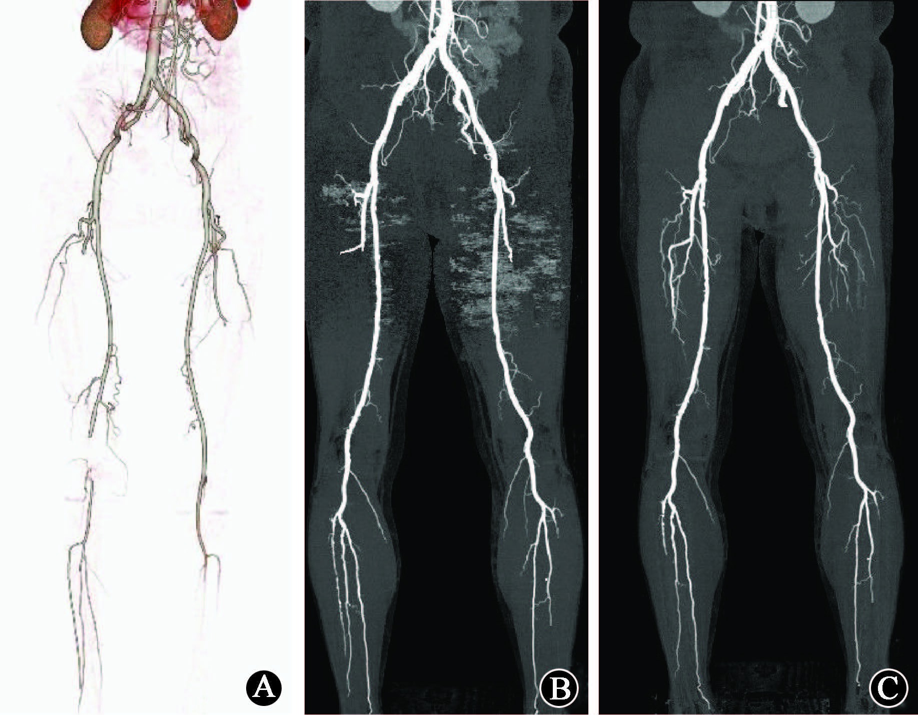
(2)两组主观图像质量评价:在FBP重建条件下,对照组和试验组主观图像质量评分差异无统计学意义(z= -0.57,P>0.05)。在100 kV条件下,AIDR-3D重建组主观图像质量评分高于FBP重建组(z= -3.25,P<0.05,图 3)。同样,100 kV和AIDR-3D重建组主观图像质量评分高于120 kV和FBP重建组(z= -3.53,P<0.05,表 2)。

|
图 3 下肢动脉CTA图 A.120 kV+FBP的VR图; B.100 kV+FBP的MIP图; C. 100 kV+AIDR-3D的MIP图 Figure 3 CTA images of lower extremity artery A. Volume rendered image using 120 kV and FBP; B.MIP image using 100 kV and FBP; C. MIP image using 100 kV and AIDR-3D |
讨论
由于辐射剂量与管电压的平方成正比,现阶段使用低管电压是降低患者辐射剂量的有效方法[5, 15]。Oca Pernas等[15]使用80 kV管电压行下肢动脉CTA检查并与100 kV管电压扫描比较,发现降低了近50%的辐射剂量。Wintersperger等[16]使用120 kV管电压行腹-髂动脉CTA检查,并与100 kV管电压比较,减少了33.7%的辐射剂量,但SNR、CNR及图像质量无影响。本研究中,试验组辐射剂量较对照组降低了54%,在同等管电压条件下,本研究DLP较低,原因可能是本研究采用了自动毫安秒技术和320排CT的大螺距所致。Leipsic等[17]认为,由于管电压的降低,增加了X射线与被检查体的光电效应,使不同密度组织间的吸收差别增大,提高了强化血管的CT值,也增加了对比度,但是降低管电压可使X射线的穿透力减低,造成到达探测器的X射线光子数量减少,导致重组图像的噪声增加。尤其在下腹部由于X射线穿透力的下降,光子散射及衰减明显增加了图像噪声。在低管电压情况下,增高的血管信号强度,可对低管电压引起的图像噪声增高起到一个补偿作用而不影响图像质量。在本研究中,在相同FBP重建条件下,试验组VD和噪声明显高于对照组。但由于两者的相互补偿,SNR、CNR和主观评分差异无统计学意义,与文献报道一致[16]。
迭代算法的出现,可以进一步弥补低管电压带来的噪声增高问题。AIDR-3D算法可以在低剂量扫描情况下可以通过多次的迭代和校正更新,重建出高质量和低噪声的图像,并对原始数据和重建图像都有效[10-13] 。本研究中,试验组中,经迭代重建处理后图像噪声减少了35%,SNR和CNR分别提高了37%和38%,而主观质量评分也有所提高。由于噪声的减少,SNR和CNR均有所提高,图像显示细节及分支血管的能力也得到提高,与Yoo等[11]报道一致。本研究中,100 kV AIDR-3D组图像噪声明显低于120 kV FBP重建组,而SNR、CNR和主观图像质量均增高。笔者认为在下肢动脉CTA检查中,迭代重建的补偿作用要大于管电压减低所引起的噪声增高。
由于CTA与DSA有很好的相关性,本研究的局限性在于病变的灵敏度特异性等方面缺乏DSA的对照,未能证实低剂量下肢动脉扫描在临床诊断方面的准确性,还有待于病例资料的完善和深入分析。
总之,在下肢动脉CTA检查中,100 kV联合AIDR-3D重建算法可明显提高图像质量,降低图像噪声,并且使辐射剂量大幅度减低,值得临床推广。
利益冲突 本研究接受宁波市自然基金项目(2016A610140) 和宁波市科技惠民项目(2016C51013)赞助。本人与本人家属、其他研究者,未因进行该研究而接受任何不正当的职务或财务津贴,并对研究的独立性和科学性予以保证作者贡献声明 李爱静设计研究方案,收集数据后统计并起草论文;宋爱芹分析及数据统计;郑建军、潘宇宁指导、监督试验进行,并修改论文
| [1] | Heijenbrok-Kal MH, Kock MC, Hunink MG. Lower extremity arterial disease: multidetector CT angiography meta-analysis[J]. Radiology, 2007, 245 (2) :433–439 . doi:10.1148/radiol.2451061280 |
| [2] | Napoli A, Anzidei M, Zaccagna F, et al. Peripheral arterial occlusive disease: diagnostic performance and effect on therapeutic m anagement of 64-section CT angiography[J]. Radiology, 2011, 261 (3) :976–986 . doi:10.1148/radiol.11103564 |
| [3] | Willmann JK, Baumert B, Schertler T, et al. Aortoiliac and lower extremity arteries assessed with 16-detector row CT angiography: prospective comparison with digital subtraction angiography[J]. Radiology, 2005, 236 (3) :1083–1093 . doi:10.1148/radiol.2362040895 |
| [4] | 黄强, 王剑锋, 翟仁友, 等. 无创影像检查方法诊断糖尿病下肢动脉病变的价值[J]. 中华放射学杂志, 2015,49 (1) :20–24. doi:10.3760/cma.j.issn.1005-1201.2015.01.006. Huang Q, Wang JF, Qu RY, et al. Diagnostic value of non-invasive imaging modalities to evaluate lower limb arterial disease associated with diabetes[J]. Chin J Radiol, 2015, 49 (1) :20–24 . doi:10.3760/cma.j.issn.1005-1201.2015.01.006. |
| [5] | Utsunomiya D, Oda S, Funama Y, et al. Comparison of standard and low-tube voltage MDCT angiography in patients with peripheral arterial disease[J]. Eur Radiol, 2010, 20 (11) :2758–2765 . doi:10.1007/s00330-010-1841-4 |
| [6] | Mulkens TH, Bellinck P, Baeyaert M, et al. Use of an automatic exposure control mechanism for dose optimization in multi-detector row CT examinations: clinical evaluation[J]. Radiology, 2005, 237 (1) :213–223 . doi:10.1148/radiol.2363041220 |
| [7] | Christner JA, Zavaletta VA, Eusemann CD, et al. Dose reduction in helical CT: dynamically adjustable z-axis X-ray beam collimation[J]. AJR Am J Roentgenol, 2010, 194 (1) . doi:10.2214/AJR.09.2878 |
| [8] | Goshima S, Kanematsu M, Nishibori H, et al. CT of the pancreas: comparison of anatomic structure depiction, image quality, and radiation exposure between 320-detector volumetric images and 64-detector helical images[J]. Radiology, 2011, 260 (1) :139–147 . doi:10.1148/radiol.11101459 |
| [9] | 郭濴, 葛英辉, 史大鹏, 等. 64层螺旋CT自动毫安技术对下肢动脉血管成像辐射剂量的研究[J]. 中华放射医学与防护杂志, 2009,29 (6) :624–642. doi:10.3760/cma.j.issn.0254-5098.2009.06.023. Guo Y, Ge YH, Shi DP, et al. Feasibility study of automatic tube current modulation technique of 64-slices CT for reducing radiation doses of lower extremity arterial imaging[J]. Chin J Radiol Med Prot, 2009, 29 (6) :624–642 . doi:10.3760/cma.j.issn.0254-5098.2009.06.023. |
| [10] | Tomizawa N, Nojo T, Akahane M, et al. Adaptive iterative dose reduction in coronary CT angiography using 320-row CT: assessment of radiation dose reduction and image quality[J]. J Cardiovasc Comput Tomogr, 2012 (6) :318–324 . doi:10.1016/j.jcct.2012.02.009 |
| [11] | Yoo RE, Park EA, Lee W, et al. Image quality of adaptive iterative dose reduction 3D of coronary CT angiography of 640-slice CT: comparison with filtered back-projection[J]. Int J Cardiovasc Imaging, 2013, 29 (3) :669–676 . doi:10.1007/s10554-012-0113-6 |
| [12] | Goshima S, Kanematsu M, Nishibori H, et al. Image quality and radiation exposure in CT of the pancreas: 320-MDCT with and without adaptive iterative dose reduction versus 64-MDCT[J]. Clin Radiol, 2013, 68 (11) :593–600 . doi:10.1016/j.crad.2013.05.102 |
| [13] | Gervaise A, Osemont B, Lecocq S, et al. CT image quality improvement using adaptive iterative dose reduction with wide-volume acquisition on 320-detector CT[J]. Eur Radiol, 2012, 22 (2) :295–301 . doi:10.1007/s00330-011-2271-7. |
| [14] | Saltybaeva N, Jafari ME, Hupfer M, et al. Estimates of effective dose for CT scans of the lower extremities[J]. Radiology, 2014, 273 (1) :153–159 . doi:10.1148/radiol.14132903 |
| [15] | Oca Pernas R, Delgado Sánchez-Gracián C, Tardáguila de la Fuente G, et al. Comparison of image quality and radiation dose in computed tomography angiography of the peripheral arteries using tube voltage of 80 kV versus 100 kV[J]. Radiología, 2014, 56 (6) :541–547 . doi:10.1016/j.rx.2012.06.013 |
| [16] | Wintersperger B, Jakobs T, Herzog P, et al. Aorto-iliac multidetector-row CT angiography with low kV settings: improved vessel enhancement and simultaneous reduction of radiation dose[J]. Eur Radiol, 2005, 15 (2) :334–341 . doi:10.1007/s00330-004-2575-y |
| [17] | Leipsic J, Nquyen G, Brown J, et al. A prospective evaluation of dose reduction and image quality in chest CT using adaptive statistical Iterative reconstruction[J]. AJR Am J Roentgenol, 2010, 195 (5) :1095–1099 . doi:10.2214/AJR.09.4050 |
| [18] | Mieville FA, Gudinchet F, Brunelle F, et al. Iterative reconstruction methods in two different MDCT scanners: Physical metrics and 4-alternative forced-choice detectability experiments-a phantom approach[J]. Phys Med, 2013, 29 (1) :99–110 . doi:10.1016/j.ejmp.2011.12.004 |
| [19] | May MS, Wust W, Brand M, et al. Lell MM (2011) dose reduction in abdominal computed tomography: intraindividual comparison of image quality of full-dose standard and half-dose iterative reconstructions with dual-source computed tomography[J]. Invest Radiol, 2011, 46 (7) :465–470 . doi:10.1097/RLI.0b013e31821690a1 |
| [20] | Pontana F, Duhamel A, Pagniez J, et al. Chest computed tomography using iterative reconstruction vs. filtered back projection (Part 2): image quality of low-dose CT examinations in 80 patients[J]. Eur Radiol, 2011, 21 (3) :636–643 . doi:10.1007/s00330-010-1991-4 |
 2016, Vol. 36


